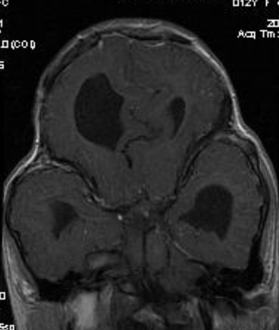
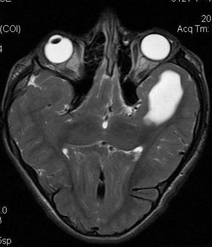
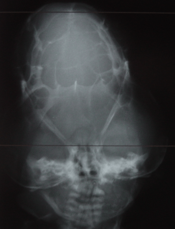
Cloverleaf skull syndrome is an abnormal configuration of the calvaria classified as craniosynostosis, consisting of premature ossification of cranial sutures. It is a deformity characterized by a remarkable enlargement of the head, with a trilobed configuration of the frontal view, resembling a three leaved clover. This abnormality occurs as a result from a severe alteration in the development of the skull, with premature synostosis of some cranial sutures, most commonly the coronal and lambdoid sutures, in association with hydrocephalus, leading to a marked bulging of the head in the region of the anterior fontanel and laterally in the temporal regions, with the typical appearance of a «cloverleaf». Syndromic and nonsyndromic presentations have been reported. Because of the anomalies both in the calvaria and in the skull base and face, this is one of the craniosynostosis currently requiring the most complex multidisciplinary approach.
The precise etiopathogenesis of this syndrome is still to be completely known, with theories involving altered membranous-osseous and/or endochondral ossification, generalized chondrodysplastic process, and a possible vascular origin associated with the abnormal osteoclastic resorption. Recently, genetic investigations have contributed to advances in the understanding of the molecular basis of some craniosynostosis syndromes, highlighting mutations in the genes FGFR1, FGFR2, FGFR3, TWIST and MSX2.
The diagnosis of such a syndrome can be made in the prenatal period by means of ultrasonography, which detects the altered cranial morphology and hydrocephalus. Traditionally, the diagnosis occurs during routine prenatal follow up at the second gestational trimester. However, with the increasing use of obstetric ultrasonography at the first gestational trimester, such alterations may be detected increasingly earlier over the gestation.
Ref: Radiol Bras. 2014 May-Jun; 47(3): 189–190.doi: 10.1590/0100-3984.2013.1681