

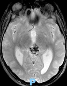
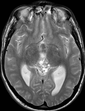
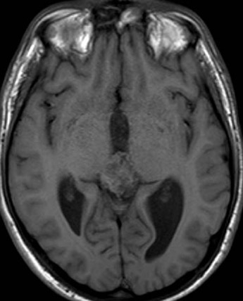

Pineal parenchyma tumour of intermediate differentiation (PPTID) accounts for up to 20 % of all pineal parenchymal tumours. They are characterised by diffuse sheets or lobules of cells similar to pineocytomas and pineoblastomas with mild to moderate nuclear atypia and low to moderate mitotic activity. PPTID are of intermediate-grade malignancy and are classified as WHO grade II or III neoplasm. The peak prevalence is during adulthood; however, they can occur at all ages and have a female predilection. The 5-year survival is 39–79 %, which is between the outcomes of pineocytoma and pineoblastoma .
No specific MRI findings distinguish PPTID from pineocytomas or pineoblastomas. Generally, PPTID are lobulated, vascular pineal region masses that can extend into adjacent structures such as the ventricles or thalami. Owing to high cellularity, PPTID are usually hyperdense on CT scans and can demonstrate peripheral exploded calcifications. On MRI, these tumours are heterogeneously hypointense on T1 weighted and heterogeneously hyperintense on T2 weighted images. Cystic areas can be seen within the tumour as well. Heterogeneous enhancement is typical. Hydrocephalus is often seen owing to mass effect on the tectum. Rarer complications include intracranial dissemination and cerebrospinal fluid spread to the spine.
References: