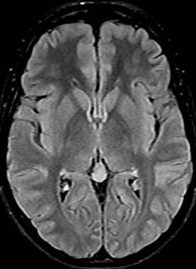
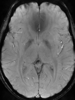
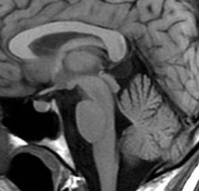
Pineal cysts are frequently asymptomatic, small sized, unilocular, benign pineal lesions which do not show size change. They are generally made of three layers: fibrocollagen layer at the outside, pineal parenchymal layer which may have calcium deposits at the middle and hypocellular glial tissue layer which may have hemosiderin inside. PCs may develop as secondary to focal degeneration of pineal gland or distension of pineal diverticulum remnant. Unilocular, smooth edged, round or ovoid shaped cysts which have homogenous interior signal feature and rim-shaped contrast-enhancement with less than 2 mm wall thickness on MRI are referred as typical PC.
Their frequency is higher in women and adults, and their sizes are not associated with gender or age. Great majority of them are isointense with CSF on T1 and T2A series. On FLAIR sequence, they are hyperintense compared to CSF, and they may be smoothly contoured, unilocular or multilocular. Typical ones may have contrast-enhancement in peripheral rim style, while multilocular ones may have septal contrast-enhancement.
Reference: