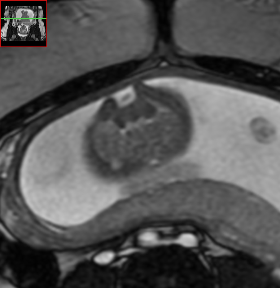
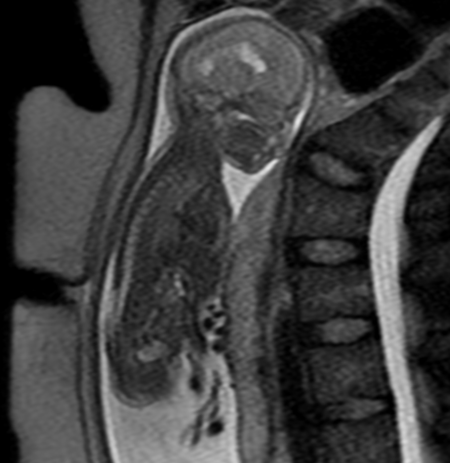
Chiari II malformation is a congenital anomaly of the brain that occurs due to incomplete development of the posterior fossa, leading to herniation of the cerebellar vermis and brainstem through the foramen magnum. Fetal MRI is a valuable imaging tool for detecting and characterizing Chiari II malformations prenatally.
On fetal MRI, Chiari II malformation typically appears as a downward displacement of the cerebellar vermis and brainstem through the foramen magnum. This results in a characteristic «banana sign,» which refers to the abnormal curvature of the brainstem and cerebellum. Additionally, there may be ventriculomegaly and aqueductal stenosis due to associated hydrocephalus.
Other findings on fetal MRI may include spinal dysraphism, such as myelomeningocele, which is present in up to 90% of cases. There may also be associated anomalies of the posterior fossa, such as absent or hypoplastic cerebellar hemispheres and vermis, and absent or hypoplastic fourth ventricle. These additional findings are important to consider in the overall management and prognosis of the affected fetus.