

Neuroepithelial cysts, also known as glioependymal cysts, originate from the area surrounding the craniospinal axis (ie, orbital, intraspinal, perispinal, sacral, and pericranial). The cysts develop from ectopic ependymal cells.
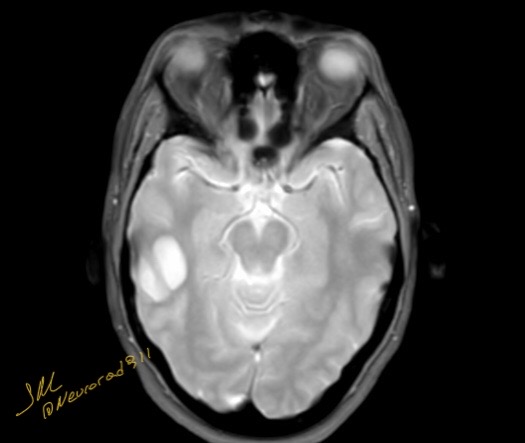
Both MRI and CT play an essential role in establishing a diagnosis, although MRI is the preferred modality for identifying neuroepithelial cysts. The cyst has smooth, rounded borders and is composed of thin, unilocular walls that contain CSF-like fluid that is identifiable on CT or MRI. CT scans generate a cystic focus with unenhanced low-density fluid, and MRI shows a cystic-filled lesion with fluid that is hypointense on T1-weighted and hyperintense on T2-weighted images. Moreover, the contents of the cyst show no restriction in the diffusion-weighted sequence, which is comparable to CSF and no surrounding edema. The mass effect caused by these cysts is minimal, even in prominent lesions, indicating their marginal growth, especially in adults. There is no enhancement after contrast media injection
Reference: