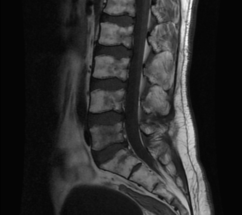
In patients with sickle cell disease (SCD), the spine can be affected in various ways, including ischemic injury, bone infarction, and vertebral collapse. MRI is a useful imaging tool to detect these changes and assess the severity of spinal involvement.
The following are some common MRI findings in SCD patients with spinal involvement:
- Vertebral compression fractures: These are common in SCD patients due to the weakened bone structure resulting from chronic anemia and ischemic injury. On MRI, vertebral compression fractures appear as wedged-shaped vertebral bodies with decreased height and abnormal signal intensity.
- Bone infarction: This is a common complication of SCD that can affect the spine. On MRI, bone infarction appears as areas of low signal intensity on T1-weighted images and high signal intensity on T2-weighted images.
- Disk degeneration: Patients with SCD are more likely to develop disk degeneration due to the chronic anemia and ischemic injury. On MRI, disk degeneration appears as reduced disk height, loss of disk hydration, and bulging or herniation of the disk.
- Vertebral collapse: In severe cases, SCD patients can develop vertebral collapse due to weakened bone structure. On MRI, vertebral collapse appears as a loss of vertebral height and increased signal intensity on T2-weighted images.
- Spinal cord compression: In rare cases, SCD patients can develop spinal cord compression due to vertebral collapse or herniated disk. On MRI, spinal cord compression appears as a narrowed spinal canal and compression of the spinal cord.
Reference: