
Vertebral haemangiomas are common vascular lesions affecting 11% of the population according to autopsy series. These lesions are usually asymptomatic and are discovered as incidental findings on multiple imaging modalities. Nevertheless, in some instances, these lesions can behave aggressively and cause pain or neurological deficit secondary to expansion of the vertebral body or paravertebral or epidural soft-tissue extension with spinal cord/nerve root compression.
Histologically, haemangiomas are predominantly composed of vascular lined spaces and non-vascular components that may include adipose tissue, smooth muscle, fibrous tissue, bone, haemosiderin and thrombus.
There are two histological subtypes of haemangioma: the cavernous type which is the most common subtype and is characterized by large sinusoidal spaces, and the capillary type which demonstrates smaller vascular channels.
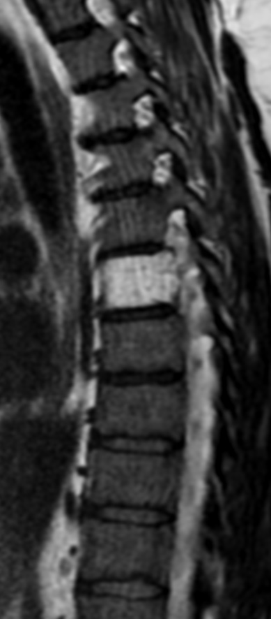
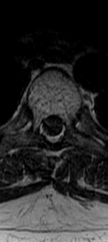
Vertebral haemangiomas classically have a coarse, vertical, trabecular pattern on radiographs which are seen on axial CT scan as punctate areas of sclerosis on axial imaging called “polka-dot” appearance or “jail-bar”, or “corduroy-cloth appearance” on sagittal or coronal reconstructions.
On MRI imaging, the signal intensity (SI) of typical asymptomatic haemangiomas is usually hyperintense on both T1 and T2 weighted sequences; however, aggressive haemangiomas have been shown to have low T2 SI related to paucity of adipose tissue.
The presence of adipose tissue in haemangiomas therefore has been suggested as a predictor of benign nature in some studies.
Laredo et al investigated the appearance of the stroma between the osseous trabeculae on vertebral haemangiomas and demonstrated absence of fat attenuation on CT in aggressive haemangiomas, in comparison to asymptomatic haemangiomas. In their study, aggressive haemangiomas showed numerous packed, thin-walled vascular cavities with absence of fatty replacement on histopathological evaluation. Ross et al and Pastushyn et al also demonstrated that the paravertebral extraosseous component of aggressive haemangiomas is hypointense on T1. In the study by Ross et al, histopathological evaluation of the extraosseous portion of the tumour showed a preponderance of angiomatous tumour vessels and fibrous tissue, with little adipose tissue. Although a few of the previous studies showed hyperintensity on STIR sequence in their published figures of aggressive haemangioma.
Reference: