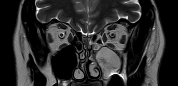
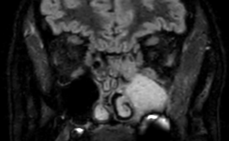
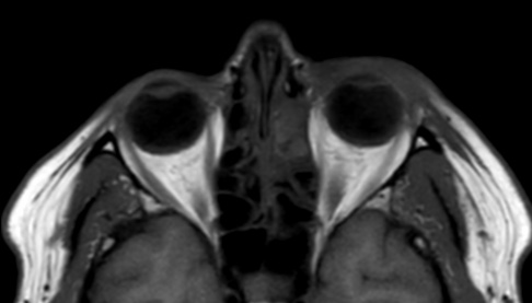

Orbital cellulitis is an infection of the soft tissues within the orbit (eye socket) of the skull. MRI is a useful imaging modality for evaluating orbital cellulitis due to its high resolution and ability to differentiate soft tissue structures.
On MRI, orbital cellulitis typically appears as a diffuse, ill-defined area of increased signal intensity on T2-weighted and fluid-attenuated inversion recovery (FLAIR) images, indicating edema and inflammation of the soft tissues within the orbit. There may also be associated enhancement of the affected tissues after intravenous administration of gadolinium-based contrast material.
In severe cases, MRI may also demonstrate abscesses or collections of pus within the orbit, which appear as areas of high signal intensity on T2-weighted and FLAIR images with associated rim enhancement after contrast administration.
Other potential findings on MRI may include proptosis, displacement of the extraocular muscles, and enlargement of the optic nerve sheath. These findings can help differentiate orbital cellulitis from other conditions that may present with similar symptoms, such as orbital tumors or Graves’ disease.
In summary, MRI findings of orbital cellulitis include a diffuse, ill-defined area of increased signal intensity on T2-weighted and FLAIR images, with associated enhancement of the affected tissues after contrast administration. Abscesses or collections of pus may also be seen in severe cases, along with proptosis, displacement of the extraocular muscles, and enlargement of the optic nerve sheath.
Reference:
DOI:10.2214/AJR.08.1838