CSF leaks can occur in patients of all ages. Most patients reveal a history of trauma or surgical procedures. However, spontaneous CSF leaks are not uncommon and are often described in middle-aged obese women with signs of intracranial hypertension.
Cerebrospinal fluid (CSF) leakage occurs when there is a tear or hole in the membranes that surround the brain and spinal cord, which causes the fluid to leak out. Traumatic CSF leaks can occur as a result of head or spinal injuries.
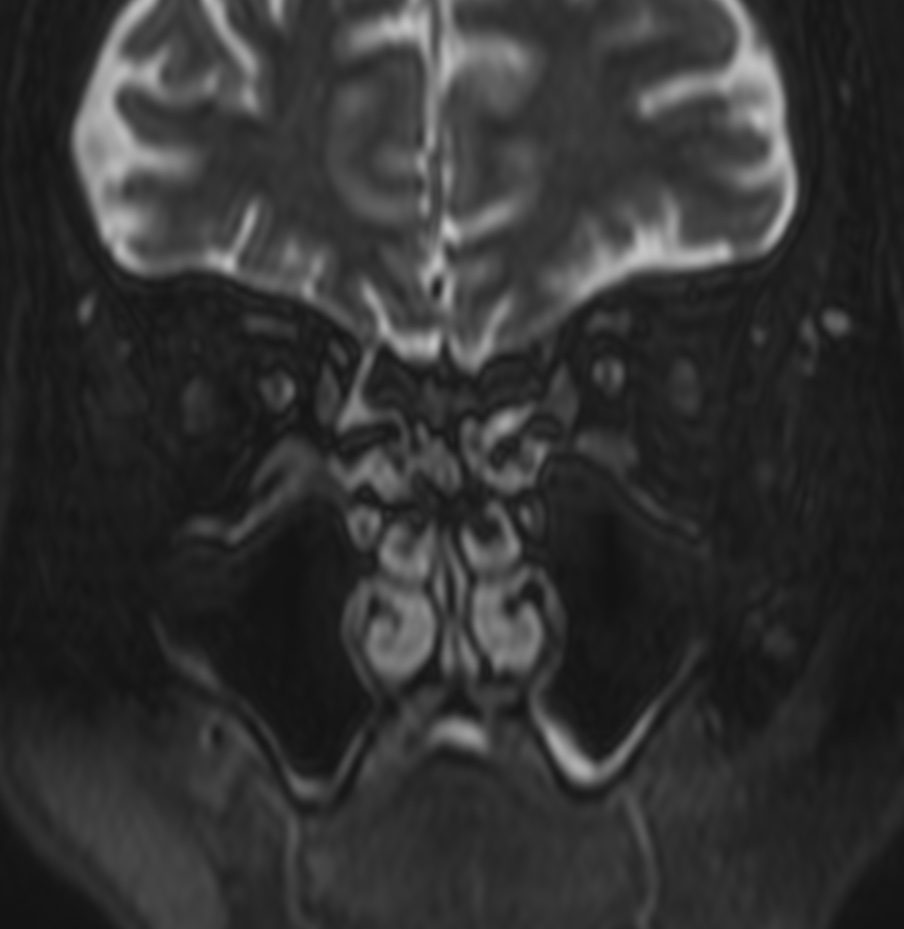
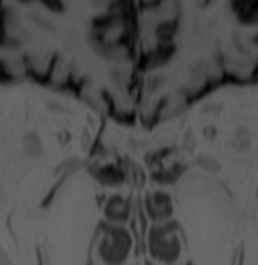
MRI (Magnetic Resonance Imaging) can be used to detect CSF leaks and identify the location of the tear or hole. The following are some of the common MRI findings associated with CSF traumatic leaks:
- Diffuse meningeal enhancement: This refers to the enhancement of the meninges, the membranes that surround the brain and spinal cord, which can be seen on contrast-enhanced MRI scans.
- Disruption of the dural sac: The dural sac is a tough, fibrous membrane that encloses the spinal cord. MRI may reveal a defect or disruption in the dural sac, which can cause CSF to leak out.
- Intracranial air: If there is a fracture in the skull base, air may enter the cranial cavity, which can be seen on MRI as hypointense areas.
- Distended pituitary gland: In some cases, a CSF leak can cause the pituitary gland to become enlarged or distended, which can be seen on MRI.
It is important to note that MRI findings alone may not be sufficient to diagnose a CSF leak. A combination of clinical symptoms, physical examination, and imaging tests may be necessary to make an accurate diagnosis.
Various imaging tests are used for identification of CSF leak. These include radionuclide cisternography, HRCT of the skull base, CT cisternography, and MR cisternography.

Reference: