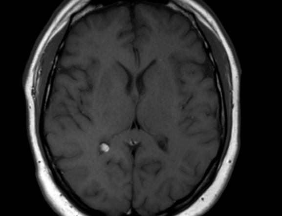

An intraventricular lipoma is a rare type of brain tumor that originates from fatty tissue within the ventricles of the brain. MRI is a preferred imaging modality for detecting intraventricular lipomas due to its high resolution and ability to differentiate soft tissue structures.
On MRI, an intraventricular lipoma appears as a well-circumscribed, homogeneous mass with low signal intensity on T1-weighted images and high signal intensity on T2-weighted images, consistent with its fatty composition. The lipoma typically appears as a well-circumscribed, lobulated mass with a smooth contour, located within the ventricular system of the brain.
In addition, the MRI may reveal any associated hydrocephalus, which is an accumulation of cerebrospinal fluid within the brain, caused by obstruction of the normal flow of fluid due to the presence of the lipoma.
In summary, MRI findings of an intraventricular lipoma include a well-circumscribed, lobulated mass with low signal intensity on T1-weighted images and high signal intensity on T2-weighted images, with associated hydrocephalus if present.
Reference: