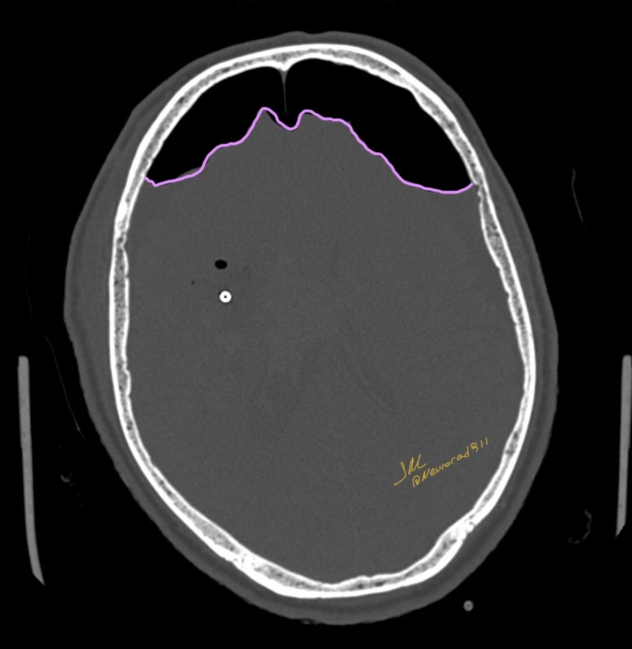
Pneumocephalus is defined as the presence of intracranial air. This is most commonly secondary to a traumatic head injury. Tension pneumocephalus presents radiologically with compression of the frontal lobes and widening of the interhemispheric space between the frontal lobes. It is often termed the Mount Fuji sign due to a perceived similarity with an iconic mountain peak in Japan.
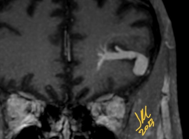
Cerebral developmental venous anomalies are the most frequently encountered cerebral vascular malformation. Most of them are asymptomatic and observed incidentally during routine CT and MRI studies. Most common vascular anomaly associated with DVA is cavernous malformation. Rarely DVAs are found in association with AV fistula. DVA associated with cerebral venous aneurysm is very rare, only few cases have been reported.
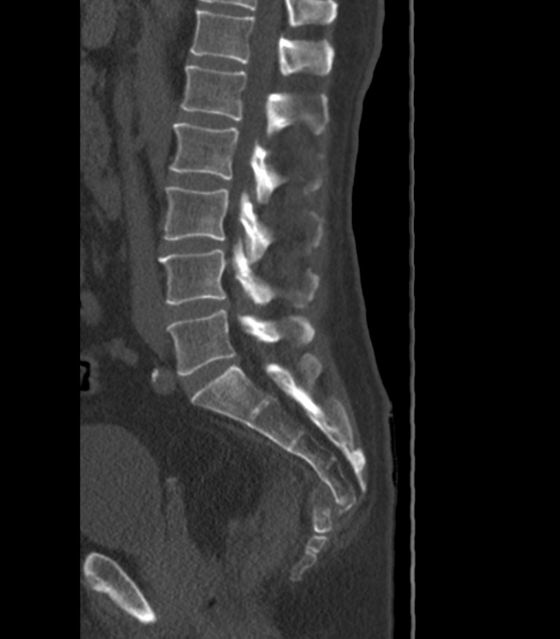
Coccydynia has a myriad of causes but is most commonly found in the posttraumatic setting. Patients often have a history of recent or remote trauma to the region and develop chronic mechanical symptoms
In these cohorts, and even in those with acute trauma and pain, fracture is rare, and static neutral radiography tend to appear normal with a similar distribution of abnormal imaging findings as in asymptomatic people.
If acute fracture is of concern, CT is typically required to make a definitive diagnosis. However, fracture may be discovered at radiography in a minority of cases.
Idiopathic and traumatic coccydynia is most commonly differentiated into categories of hypermobility, subluxation, and rigid coccyx (with or without a posterior spicule). There is a strong female predominance of coccydynia with a 4:1 female-to-male ratio. Increased body mass index (BMI) is a risk factor that is more significant in women than in men . Multiparity is also a risk factor among women . Regarding coccyx anatomy, risk factors include having a mobile sacrococcygeal joint, a more ventrally curved coccyx, and a posterior spicule formation. In men, intercoccygeal joint subluxation has been reported as a risk factor
Within the AO classification system, coccygeal fractures are classified as a subset of the sacrococcygeal fractures (classification A1)
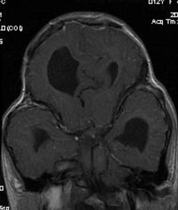
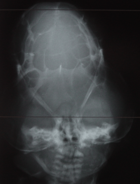
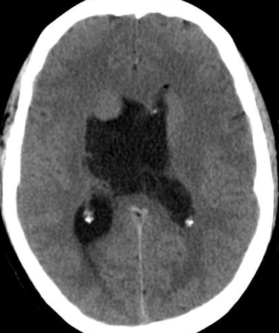
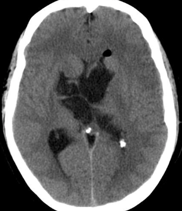
Cloverleaf skull syndrome is an abnormal configuration of the calvaria classified as craniosynostosis, consisting of premature ossification of cranial sutures. It is a deformity characterized by a remarkable enlargement of the head, with a trilobed configuration of the frontal view, resembling a three leaved clover. This abnormality occurs as a result from a severe alteration in the development of the skull, with premature synostosis of some cranial sutures, most commonly the coronal and lambdoid sutures, in association with hydrocephalus, leading to a marked bulging of the head in the region of the anterior fontanel and laterally in the temporal regions, with the typical appearance of a «cloverleaf». Syndromic and nonsyndromic presentations have been reported. Because of the anomalies both in the calvaria and in the skull base and face, this is one of the craniosynostosis currently requiring the most complex multidisciplinary approach.
The precise etiopathogenesis of this syndrome is still to be completely known, with theories involving altered membranous-osseous and/or endochondral ossification, generalized chondrodysplastic process, and a possible vascular origin associated with the abnormal osteoclastic resorption. Recently, genetic investigations have contributed to advances in the understanding of the molecular basis of some craniosynostosis syndromes, highlighting mutations in the genes FGFR1, FGFR2, FGFR3, TWIST and MSX2.
The diagnosis of such a syndrome can be made in the prenatal period by means of ultrasonography, which detects the altered cranial morphology and hydrocephalus. Traditionally, the diagnosis occurs during routine prenatal follow up at the second gestational trimester. However, with the increasing use of obstetric ultrasonography at the first gestational trimester, such alterations may be detected increasingly earlier over the gestation.
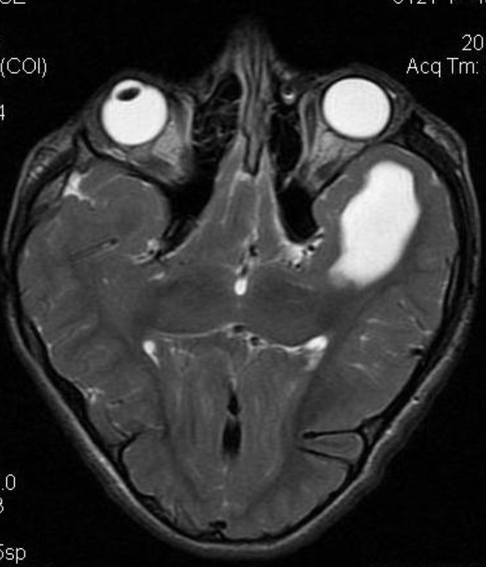
Epidermoid cysts, are rare benign tumors developed from ectodermic inclusions. They usually sit at the ponto cerebellar angle, the para-sellar region and the temporal fossa. Their location at the intraventricular is very rare. Most of the lateral intraventricular epidermoids are silent and/or do not produce hydrocephalus.