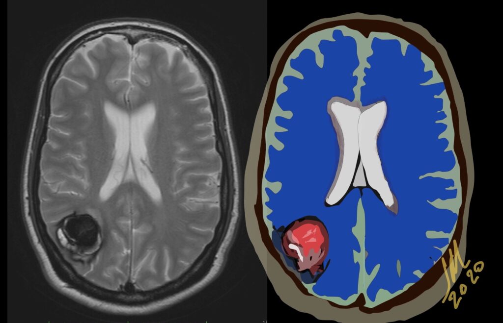
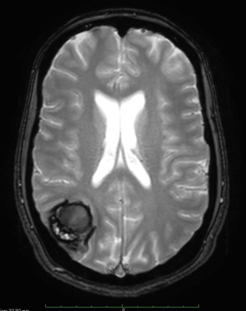
Type V indicate parts of the actual CCMs that are visible in the center of the hemorrhage; however, the CCM is not fully distinguishable from hemorrhage. Type 5 has a high hemorrhage risk of 24%
Hemorrhagic events particularly those found in relatively young patients ought to be characterized further, and cavernous angioma should be considered among the possible etiologies. Also because of their propensity to act as an epileptogenic focus CMs should always be considered in the workup of a patient with a seizure disorder, especially if the patient is aged 20-40 years.
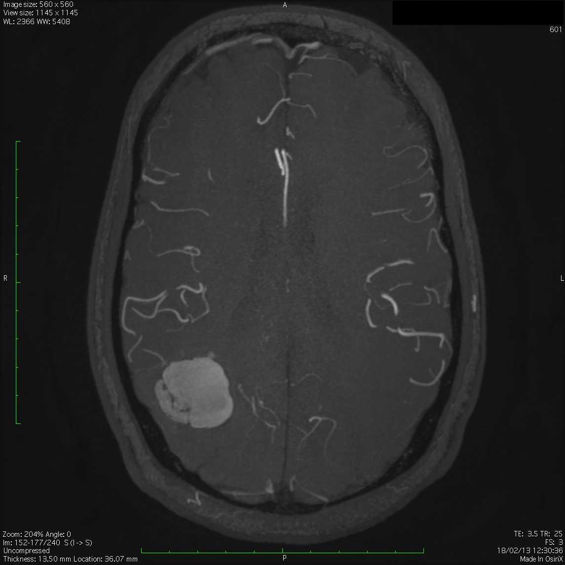
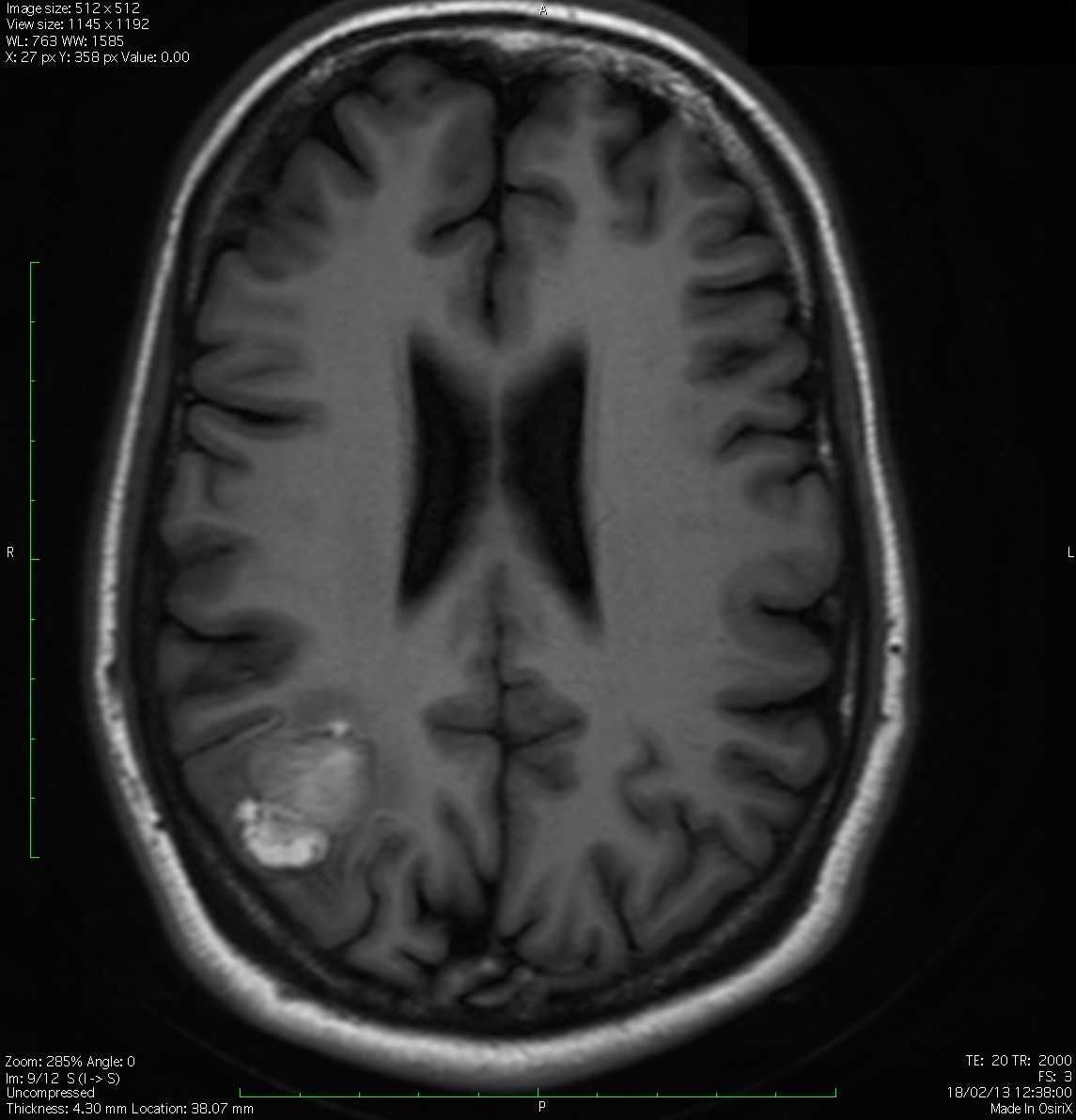
If a recent bleed has occurred then surrounding edema or mass effect may be present. The lesions show a variable pattern of enhancement. Contrast-enhanced images help delineate any potential associated developmental venous anomalies. This is critical for preoperative surgical planning as the un-deliberate resection of DVAs may compromise normal cortical venous drainage and thus lead to brain venous infarction. Angiographically these lesions are occult and usually show nonspecific findings like capillary blush or early draining vein