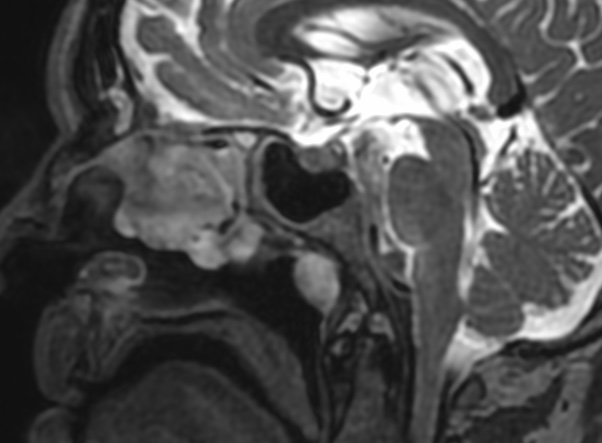
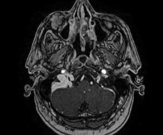
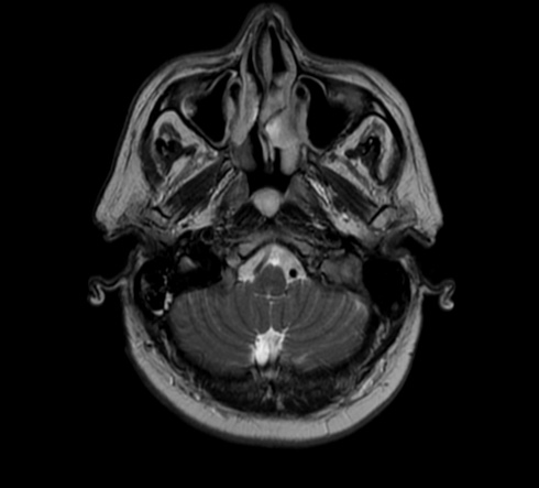
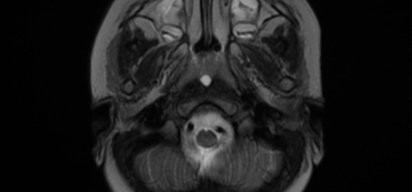
The Thornwaldt cyst is a benign cystic lesion that develops in the posterior medial wall of the nasopharynx; its incidence is 4% on autopsy samples, however an incidence of 0.2 to 5% has been observed on routine brain and cervical MRI.
The etiopathogeny remains controversial, some assume that it is a relic of the notochord (embryonic tissues from which vertebrae are formed), while others evoke an iatrogenic occlusion of normal structure after adenoidectomy or chronic inflammation
The Tornwaldt cyst is most often asymptomatic; however, an increase in its volume or inflammation may cause occipital headache, nasal obstruction with persistent discharge, bad breath, cervical myalgia, eustachian tube dysfunction, and sometimes infections of the middle ear
MRI remains the reference examination to identify this lesion, with a hypo or a T1 signal (depending on the protein content of the cyst) and a hypersignal T2
The main differential diagnoses are: Rathke’s pocket cyst, meningocele, meningoencephalocele and necrotic nasopharyngeal tumors
Reference: