
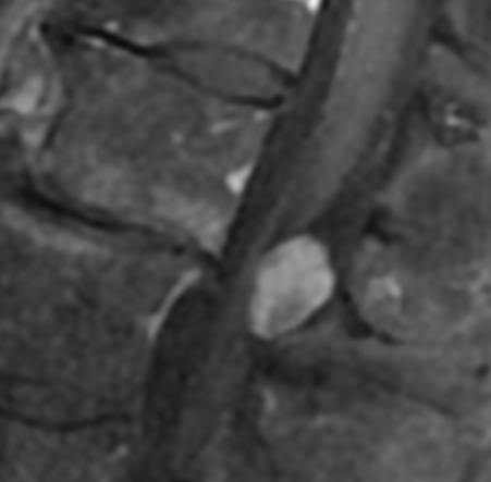
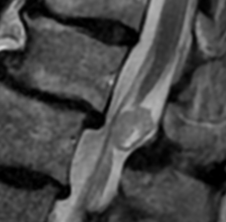
Spinal meningiomas, which are characteristically intradural and extramedullary, are relatively rare and account for less than 10% of all meningiomas and 25% of all spinal cord tumors.
Spinal meningiomas are intradural extramedullary lesions, usually benign, that are most commonly thoracic and posterolateral in location. Accounting for ~25% of spinal tumors, they are the second most common tumor in the intradural extramedullary location, second only to tumors of the nerve sheath. Most occur in middle-aged or older adults, with female predominance
Clinically, manifestations in patients with a meningioma of the lumbar spine depend on the tumor location, in relation to the spinal cord transection and nerve roots. Neurological symptoms include back or radicular pain, motor weakness, sensory disturbances, and urinary or fecal incontinence as a late finding.
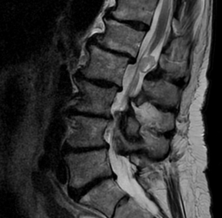
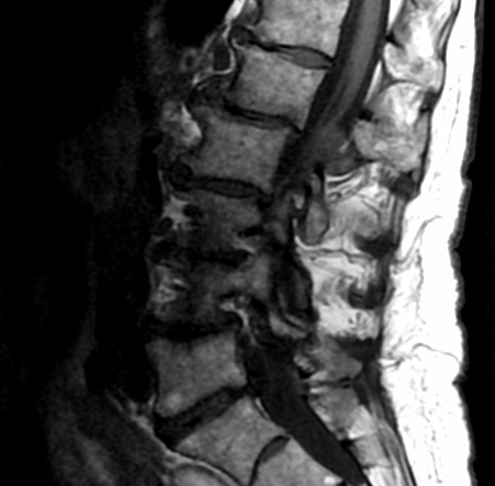
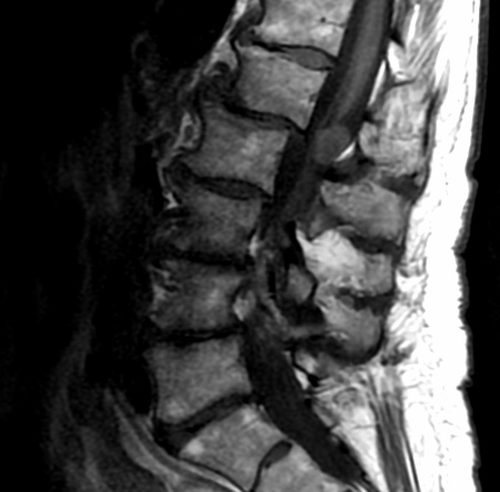
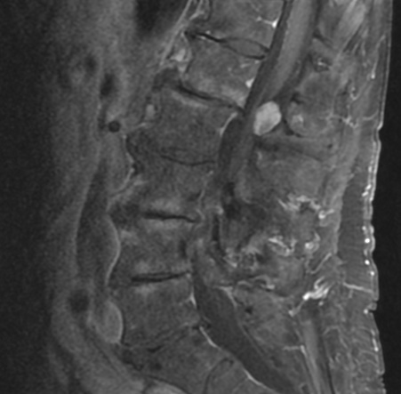
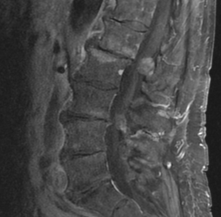
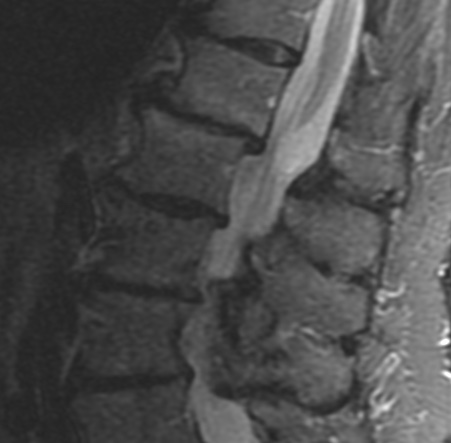
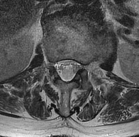
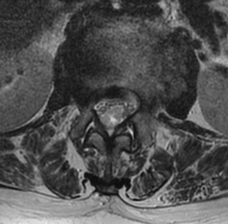
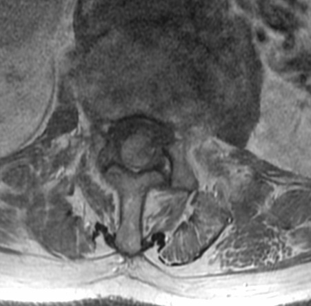
On MRI, characteristics of spinal meningiomas include well-circumscribed, broad-based dural attachment and/or a dural tail sign, which are similar signal characteristics to typical intracranial meningiomas. Spinal meningiomas typically demonstrate isointensity to slightly hypointensity on T1-weighted images, isointensity to slightly hyperintensity on T2-weighted images, and moderate homogeneous enhancement on T1-weighted images with gadolinium enhancement. Densely calcified meningiomas are sometimes hypointense on T1 and T2 images, and show only minimal contrast enhancement.
These radiological characteristics are usually nonspecific. It is difficult for physicians to differentiate benign or malignant tumors simply on images. The “dural tail sign”, for example, can be seen in intradural-extramedullary meningiomas but is also found in metastatic tumors and lymphomas.

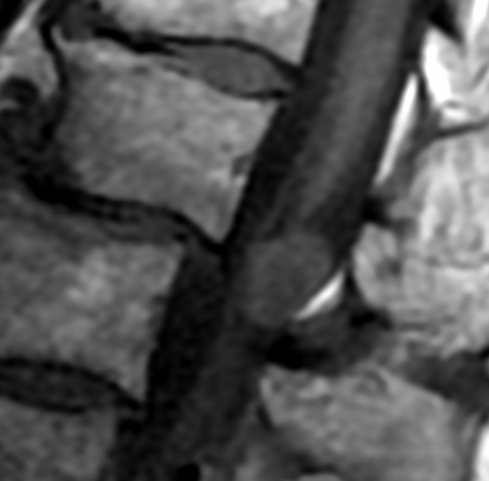

A meningioma with intradural and extradural components occasionally mimics a nerve sheath tumor and a nerve sheath tumor with a predominant intradural component may mimic a meningioma; however, nerve sheath tumors are usually hyperintense on T2, lumbar and ventral in location, rarely calcified, more common in men, and do not have a dural tail
Reference:
Yeo Y, Park C, Lee JW, et al. Magnetic resonance imaging spectrum of spinal meningioma. Clin Imaging 2019; 55:100-106
doi: 10.1016/j.ejrad.2006.04.003