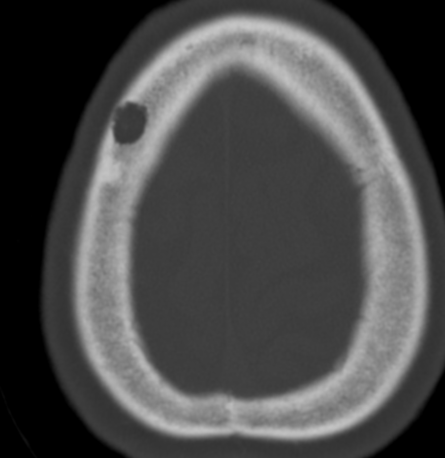
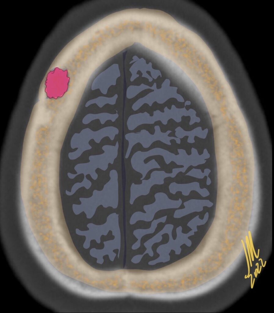
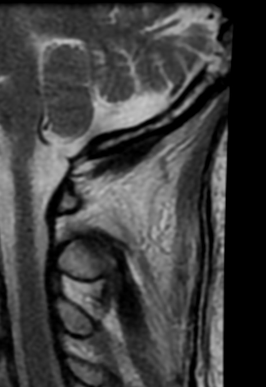
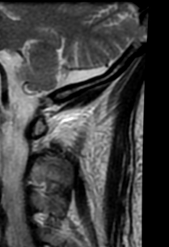
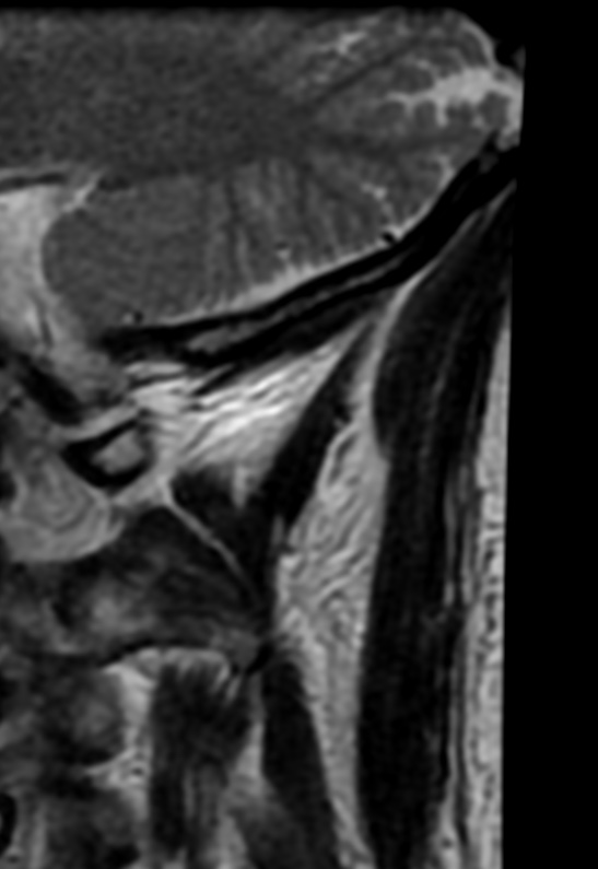
Calvarial lesions are uncommon and may constitute a diagnostic challenge. Pertinent clinical information such as patient age and history of trauma or underlying systemic disease should always be taken into consideration.
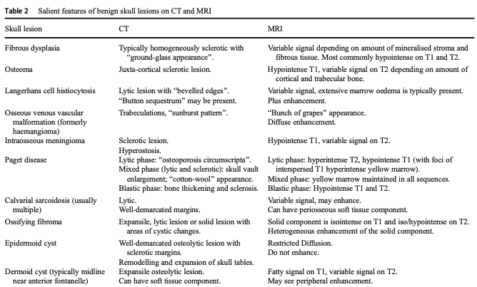
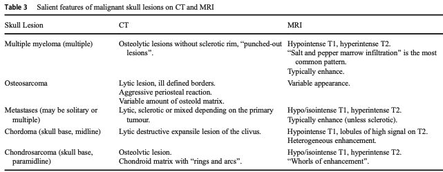
Among all imaging features, the extent, multiplicity, and attenuation of a calvarial lesion, in combination with knowledge of the patient’s age, are enough to narrow the differential diagnosis-
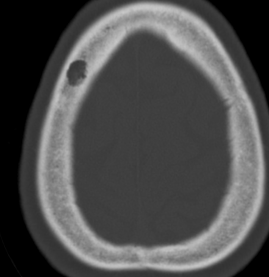

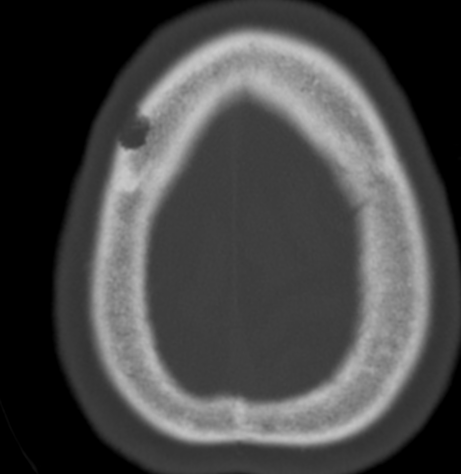
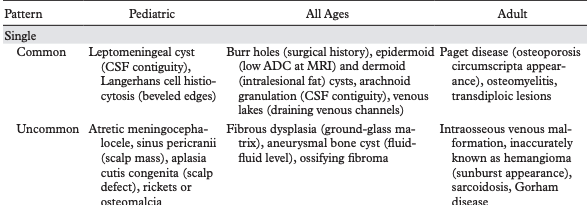
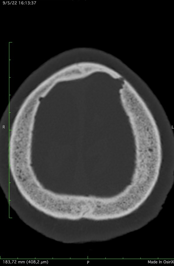
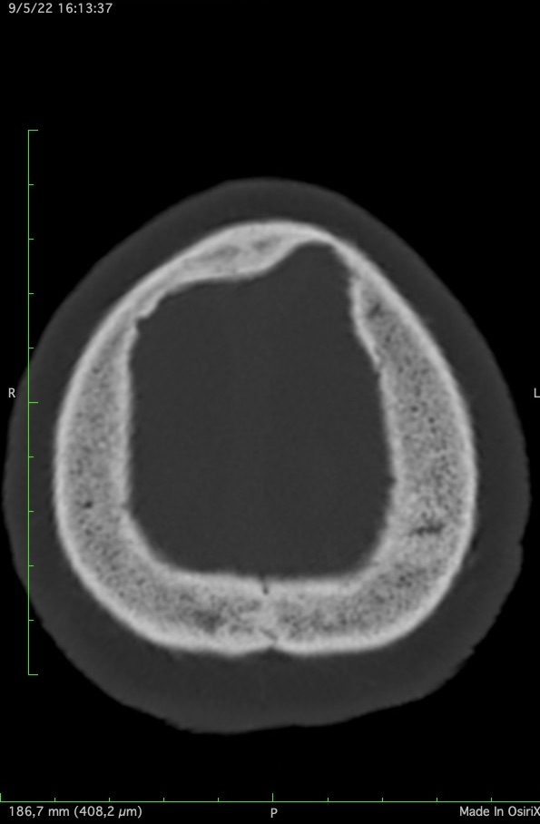
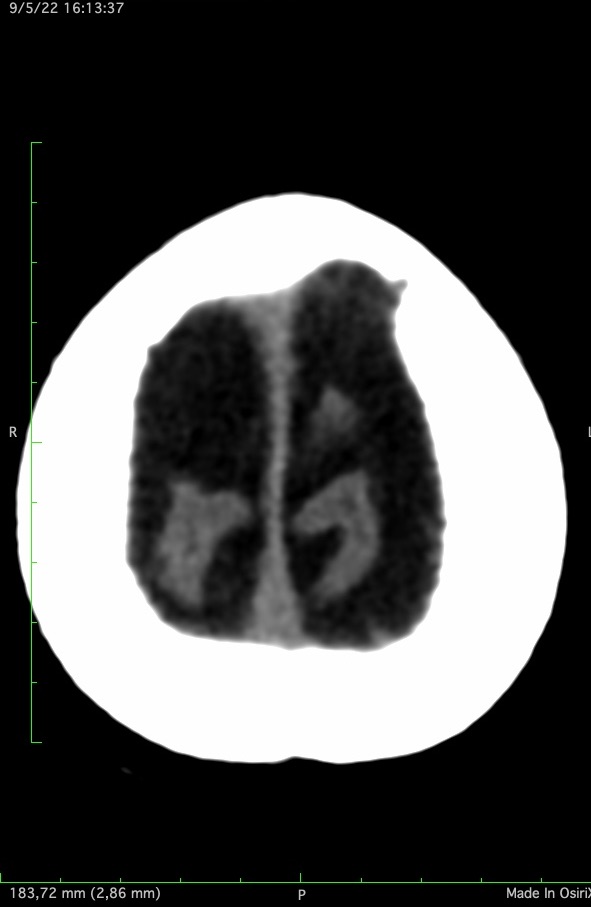
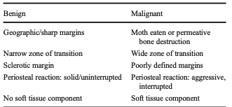
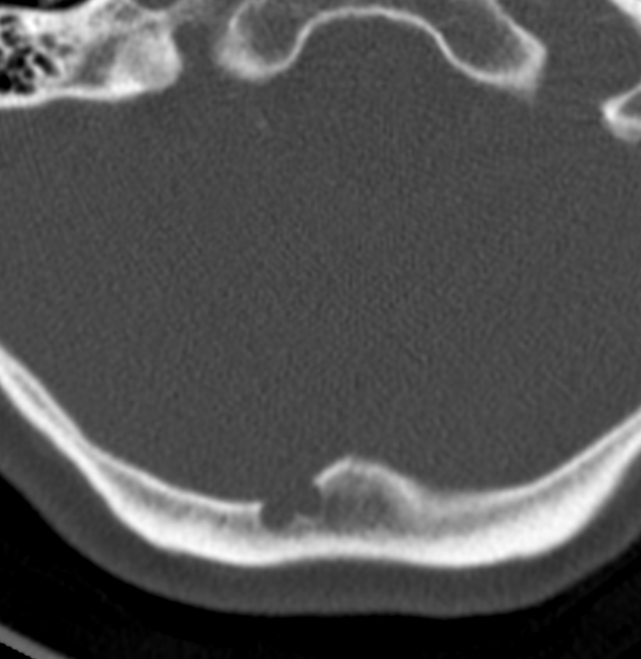
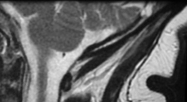
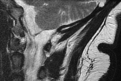
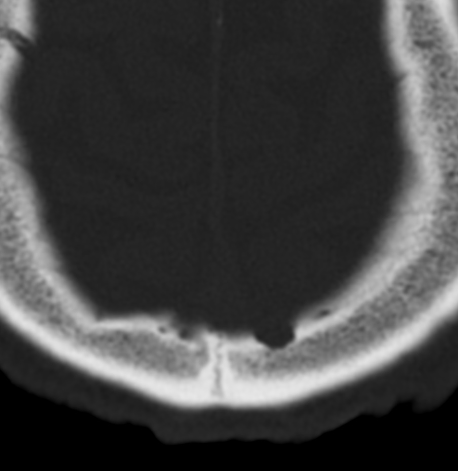
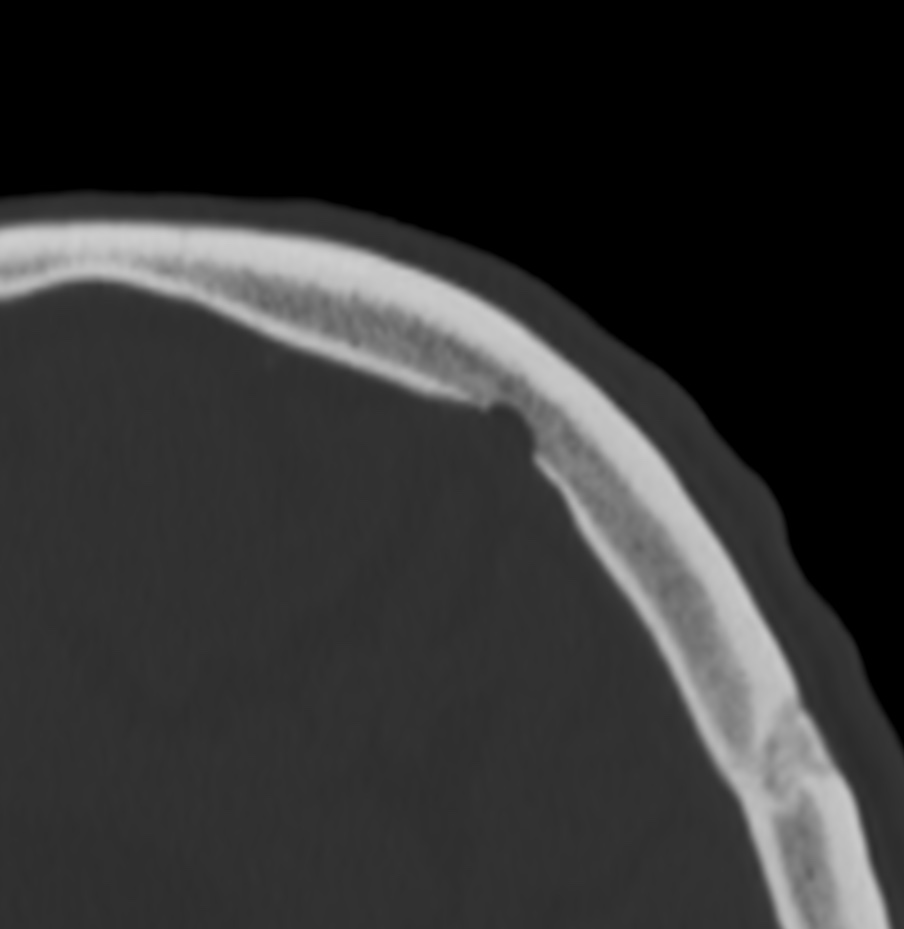
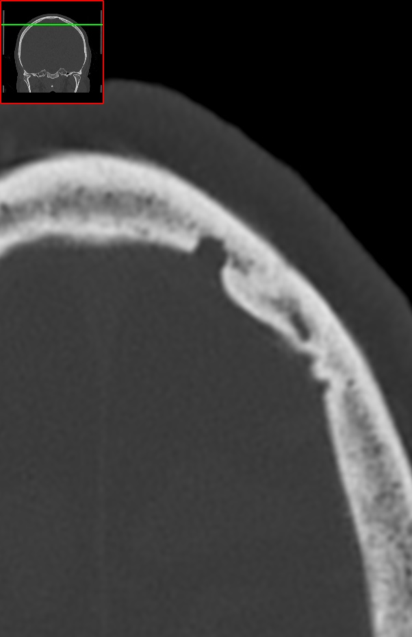
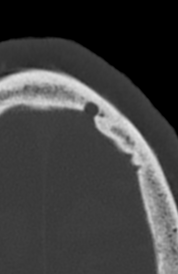
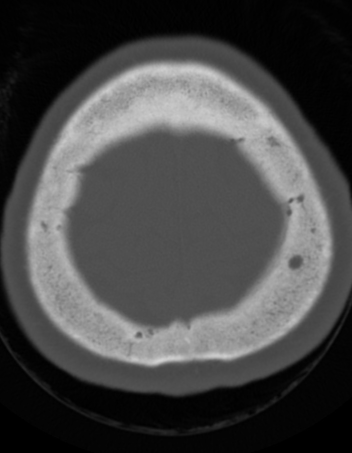
Recognition of benign and malignant imaging features is important for the radiological diagnosis. In general, benign tumours have well-defined borders with a narrow transition zone; sclerotic margins are frequently present. On the other hand, malignant tumours have poorly defined margins, a wide transition zone, aggressive periosteal reaction and often have a soft tissue component; these lesions cause dramatic bony destruction with intracranial or extracranial extension. Skull lesions can be lytic or sclerotic, single or multiple with varied composition; they may arise from osteogenic, chondrogenic, fibrogenic, vascular and/or other elements of bone.
Reference: