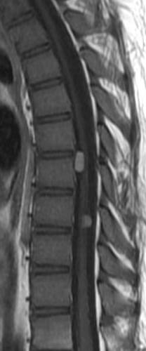
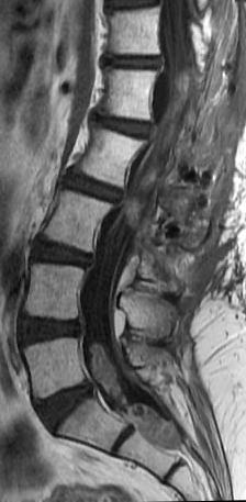
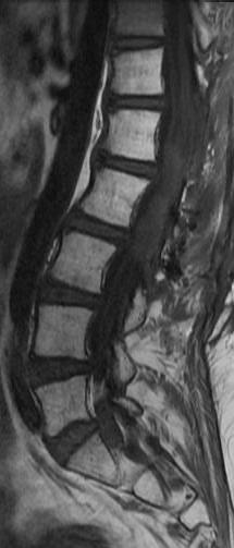
Myxopapillary ependymomas are highly vascular neoplasms characterized by abundant supporting fibrous connective tissue stroma, mucinous degeneration of this supporting stroma, and mucin secretion by the tumor cells.
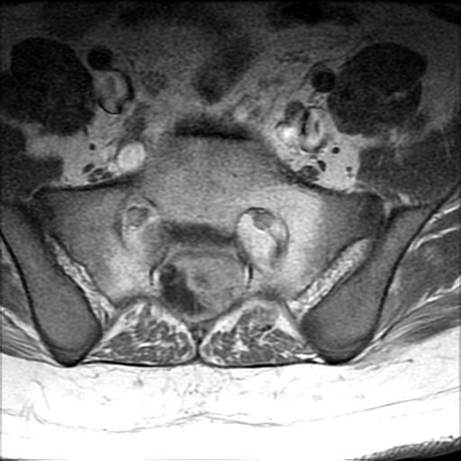
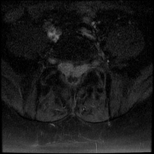
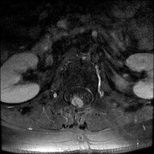
Multiple tumors have been reported in 1 4%-43% of patients and may be due to dissemination of tumor in the spinal subarachnoid space. Certain sacral and presacral lesions behave aggressively and
metastasize to the lymph nodes, lung, and bone . On the other hand, recurrence is rare following complete excision of well-circumscribed lesions.
Ependymomas of the brain frequently calcify, calcification is extremely unusual in spinal ependymoma.
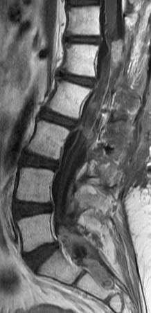
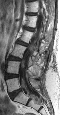
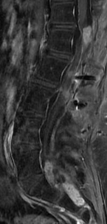
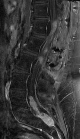
Bulky, obstructive lesions are associated with a high protein content of the CSF that may obscure differentiation of the tumor from the CSF. The signal intensity of the CSF can also be affected by subarachnoid hemorrhage. Almost all ependymomas enhance intensely after administration of
contrast material. Contrast-enhanced imaging is useful in differentiating the tumor from the spinal cord, defining intratumoral cysts, and identifying intradural metastases.
Reference: