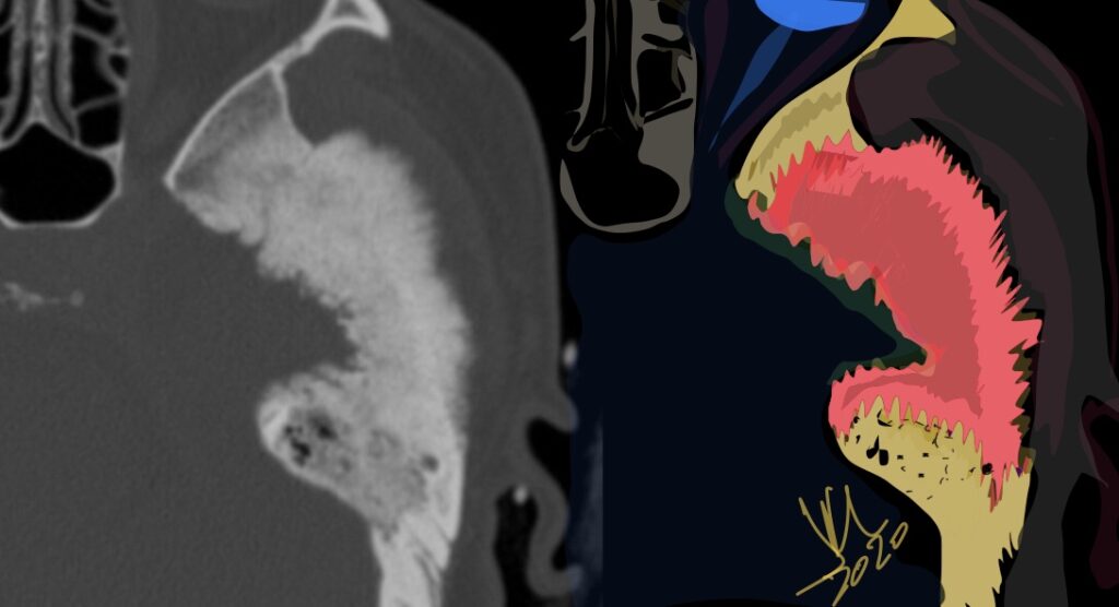
Meningioma primary to the tegmen tympani arise from the floor of the middle cranial fossa and spread inferomedially into the middle ear cavity.
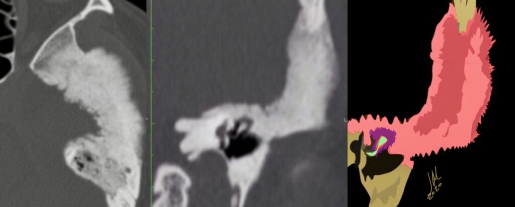
CT features included thickening of the tegmen tympani, The internal trabecular architecture of the involved bone is preserved (trabecular hyperostosis). The inner margin of the involved calvaria along the lateral aspect of the middle cranial fossa is irregular. A middle ear cavity soft-tissue mass resulted in ossicular encasement. There is no ossicular erosion or destruction. Facial nerve canal encroachment is present.
Tegmen tympani meningioma bone changes on CT can be confused with fibrous dysplasia of this area. Preservation of internal trabecular architecture distinguishes it from the ground-glass attenuation seen in typical fibrous dysplasia.
The other dominant consideration for tegmen tympani meningioma on CT is cholesteatoma; however, on CT, meningiomas lack the bone and ossicular destructive changes. These all lack the characteristic tegmen tympani thickening on CT and dural enhancement along the floor of the middle cranial fossa seen with meningioma on enhanced MR imaging. Enlargement of the facial canal typical of schwannoma is not seen with meningioma. Facial nerve hemangioma causes amorphous honeycomb bone changes on CT in larger lesions, which differ from the trabecular hyperostosis of tegmen tympani meningioma. Ossifying variants of hemangioma may have spicules of lamellar bone, which could more easily mimic meningioma on CT; however, most facial nerve hemangiomas are primarily centered in the geniculate fossa. This location should help distinguish them from meningioma