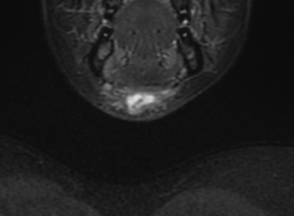
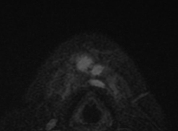

Presence of internal high attenuation, internal debris, and septations generally correlates with prior infection. In active or recent infection, patients may complain of tenderness at the site of a rapidly growing neck mass. Subsequent imaging reveals a thick-walled cyst with rim enhancement and inflammatory changes of the surrounding subcutaneous tissues.
Reference:
Patel, S., Bhatt, A.A. Thyroglossal duct pathology and mimics. Insights Imaging 10, 12 (2019). https://doi.org/10.1186/s13244-019-0694-x