
Hypoxic-ischemic encephalopathy (HIE) is the result of decreased global perfusion or oxygenation. The distribution of HIE varies according to the duration, degree, and abruptness of the hypoxic and/or ischemic insults, basal blood low, and metabolic activity in the areas of ischemia, temperature, and serum glucose levels. Layers 3, 4, and 5 of the cortex, watershed zones, and the hippocampi (cornu ammonis 1 zone) are sensitive to ischemia. DW hyperintensity throughout the cerebral cortex suggests devastating diffuse hypoxic-ischemic necrosis, whereas a pattern of basal ganglia or thalamus suggests primary hypoxic injury or mild HIE. Imaging Features include; Symmetric T2/FLAIR hyperintensity in deep gray nuclei and cortex.
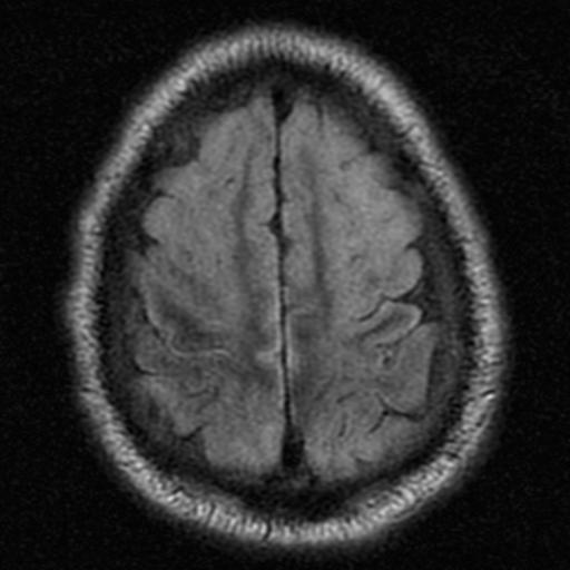
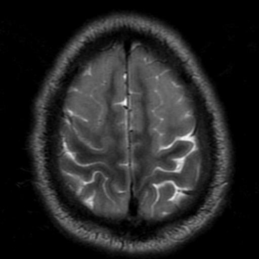
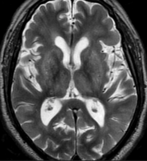
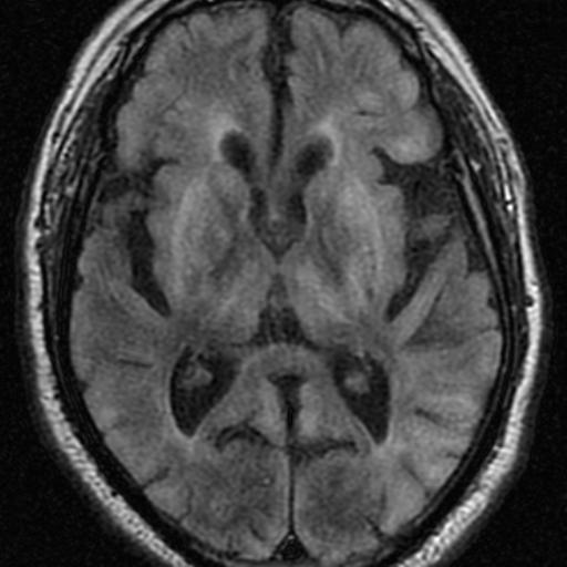
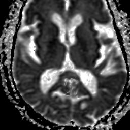
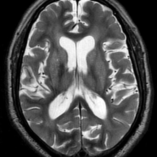
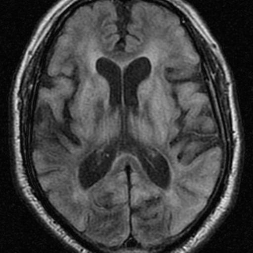
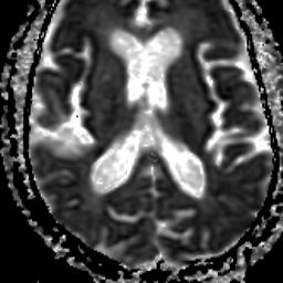
The location of the injury correlates with the severity; mild to severe> Watershed zone infarcts, Gray matter, basal ganglia, sensorimotor and visual cortex, cerebellum( in older patients)and hippocampi.
Reference:
Diffusion-Weighted MR Imaging of the Brain Moritani . Ekholm . Westesson. P- 172