Drusen are a rare inherited pathology , usually bilateral and asymptomatic, but they can lead to a visual field defect or to migraine-like headaches.
Drusen are classified into two categories: retinal and optic disc.
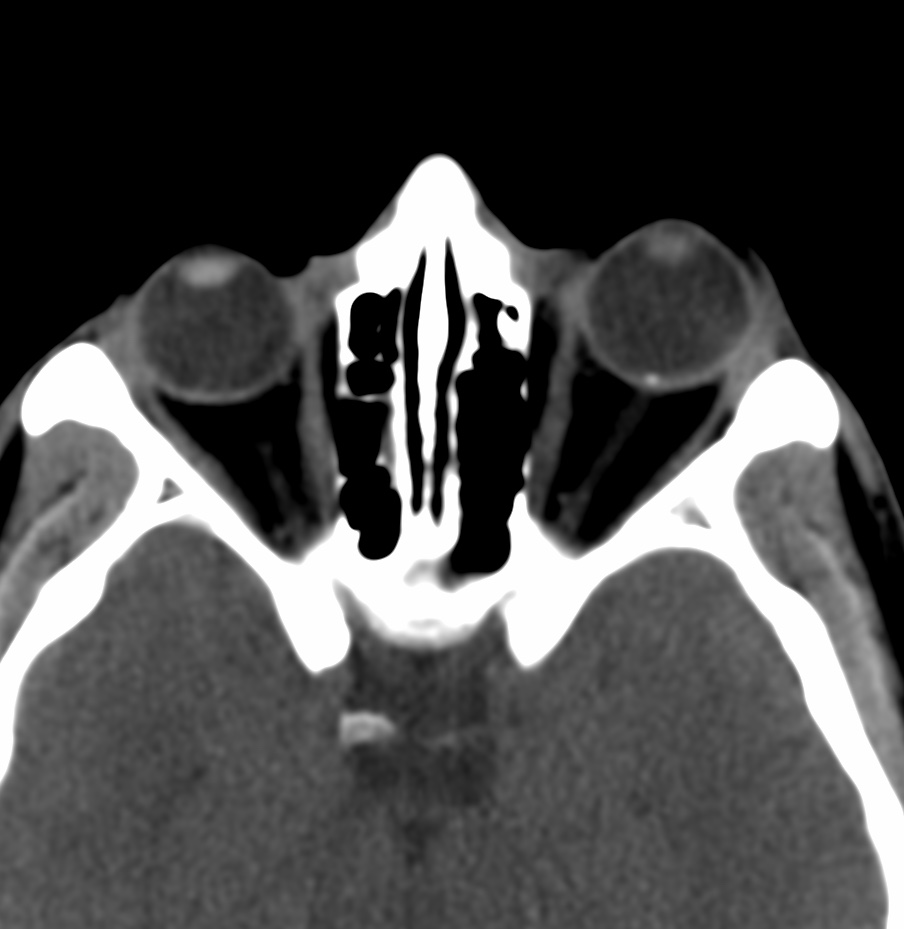
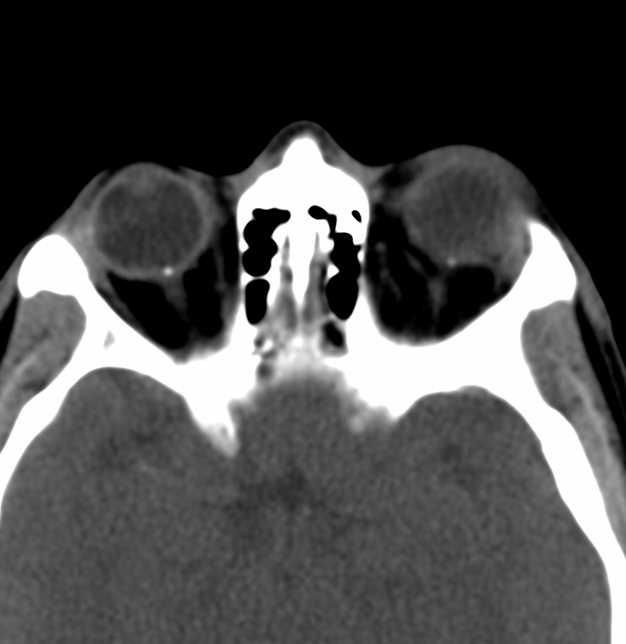
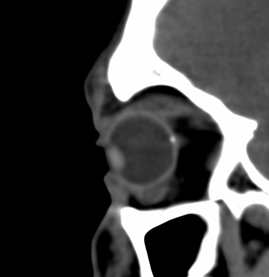
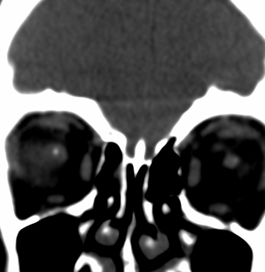
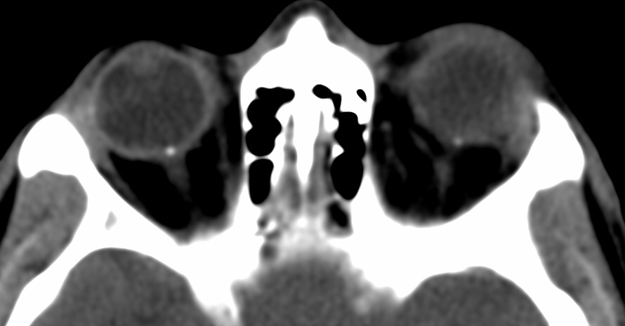
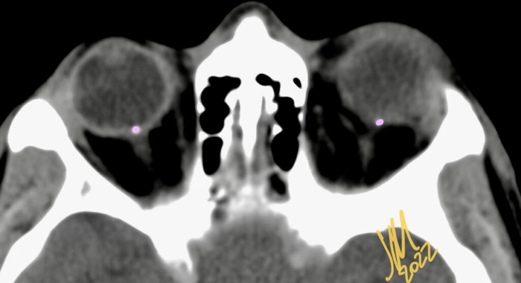
A benign calcification at the optic nerve head junction affects 0.25%-2.0% of the population and is more prevalent in White people. The condition is a result of acellular calcific deposition within the optic nerve that may mimic papilledema on fundoscopic exam, thus representing a diagnostic challenge for clinicians.
Reference:
Kumaev B, Soule E, Rao D, Fiester P. Optic Disc Drusen. Appl Radiol. 2020;49(6):54-55.
DOI: 10.1594/EURORAD/CASE.1595