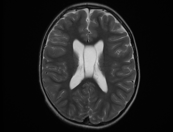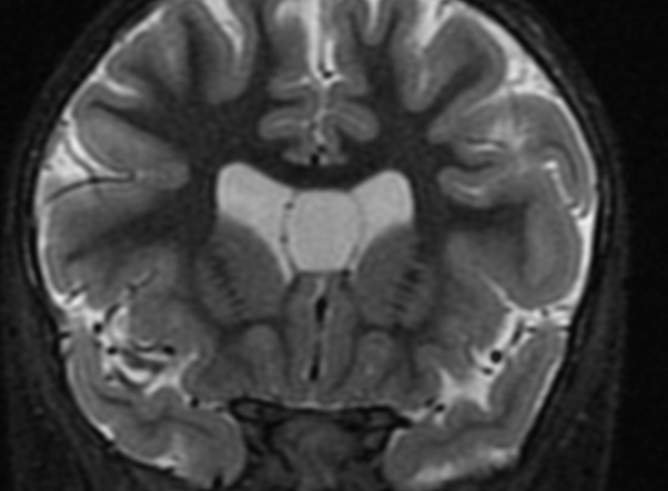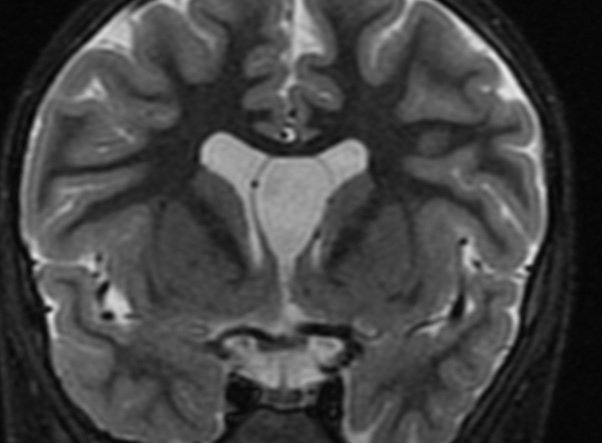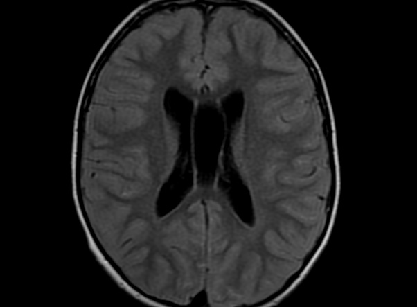


The septum pellucidum is a structure that is marginated by the corpus callosum and body of the fornix. It is composed of white matter leaves along the medial walls of the lateral ventricles and is lined by ependyma along its ventricular surfaces . The entire space between the leaves of the septi pellucidi is the cavum septi pellucidi et vergae, with the space anterior to either the foramina of Monro or an arbitrary vertical plane formed by the forniceal columns being the CSP and the space posterior being the cavum vergae.
When the two leaves fail to fuse and form the septum pellucidum, a number of postnatal anatomic variants can result including the CSP, cavum vergae, and cavum veli interpositi. The CSP and the cavum vergae usually freely communicate with one another. Usually a cavum vergae is seen in association with a CSP, although it can also occasionally be seen in isolation . In general, persistence of the CSP in postnatal life is considered a normal variant.
The leaves of the septi pellucidi begin to close at approximately 6 months’ gestational age from back to front. Nearly all term infants have closure of the cavum vergae and the majority of infants 3–6 months old have closure of the cavum septi as well . The cavum sometimes persists into adulthood as a normal variant, typically small, measuring less than 4 mm in transverse diameter, in healthy individuals . The terminology can be confusing. In general, when the two leaves are separated, this may be referred to as the “cavum septi pellucidi” or “CSP”; when the leaves are fused to form a single structure, it is referred to as the “septum pellucidum”
Reference: https://www.ajronline.org/doi/10.2214/AJR.17.19219