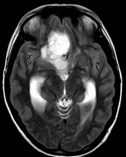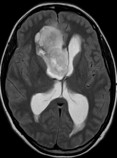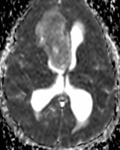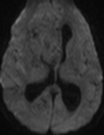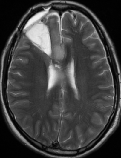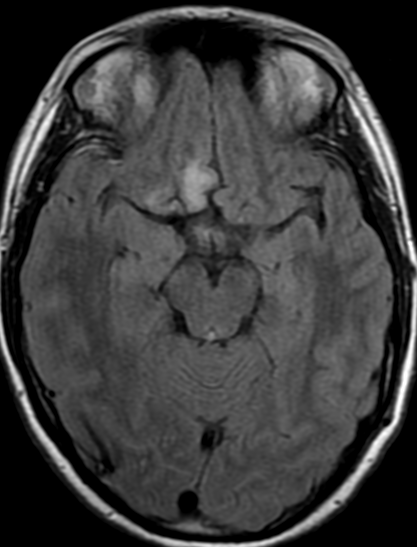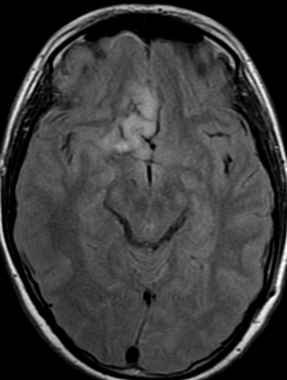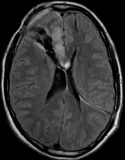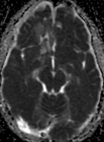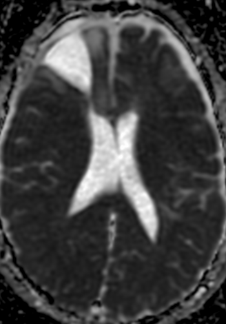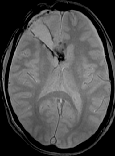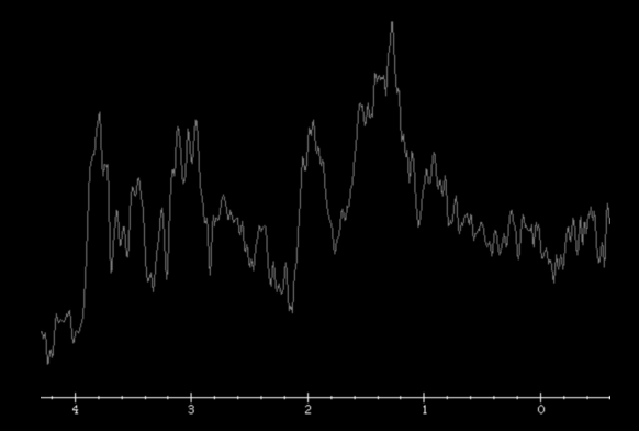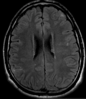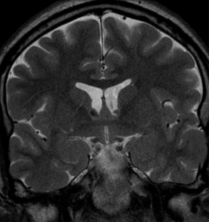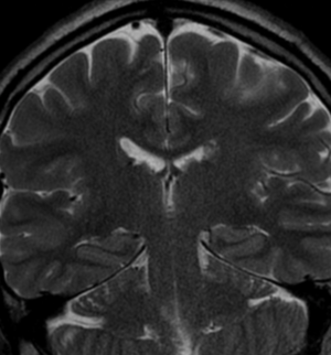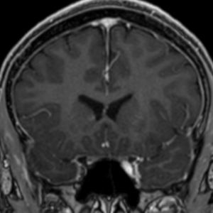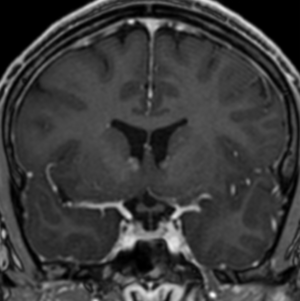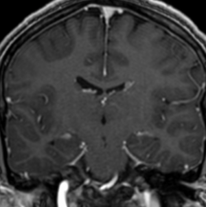
Pseudoprogression Astrocytoma-G3-Idh-wilde

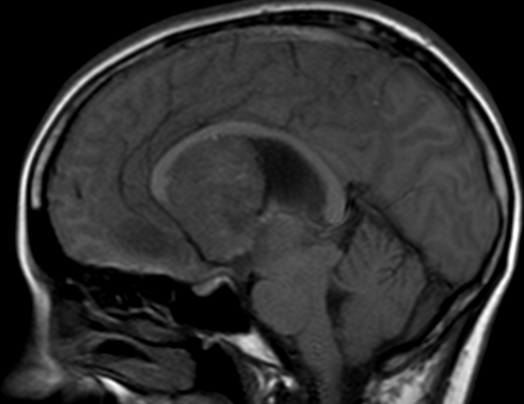
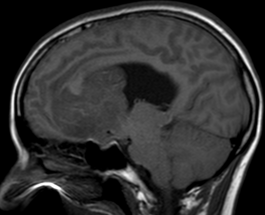
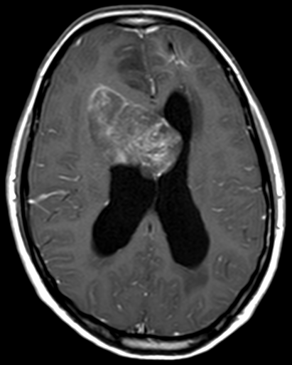
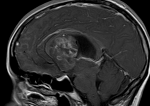
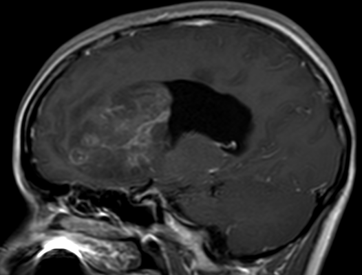
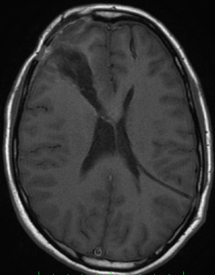
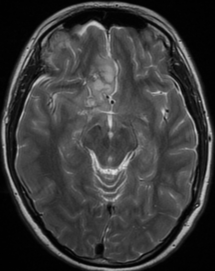
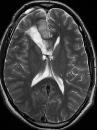
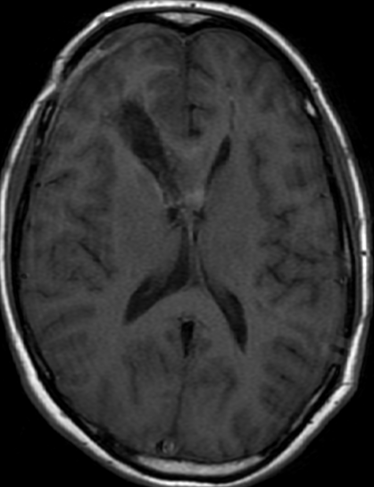
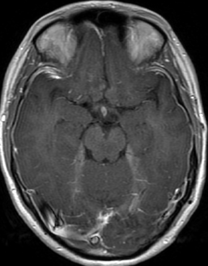
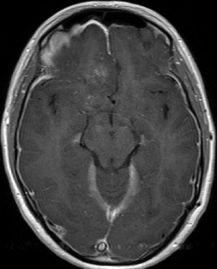
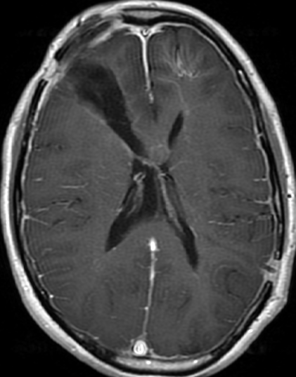
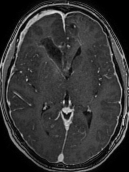
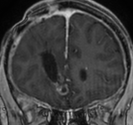
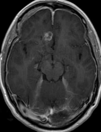
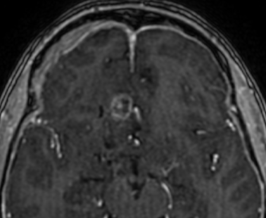
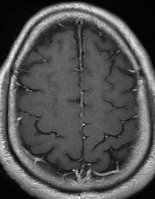
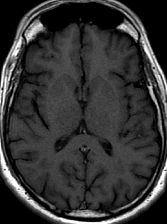
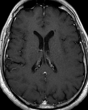
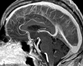
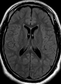
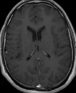
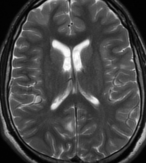
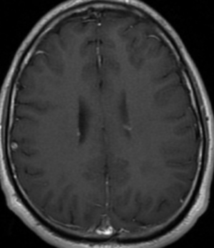
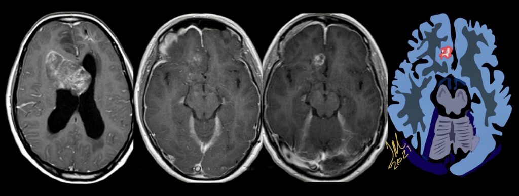
34F. First MRI. 2 months after surgery and 5 months later

Pseudoprogression is a subacute treatment-related effect with MRI features mimicking those of tumor progression. Patients can present with an increase in contrast enhancement and peritumoral edema at MRI. The diagnosis of pseudoprogression is typically made retrospectively on the basis of spontaneous improvement or stabilization of imaging findings without intervention.

Pseudoprogression typically develops in the setting of combined RT and temozolomide therapy for high-grade or low-grade glioma. but is also observed with immune checkpoint inhibitors in combination with RT for brain metastases and in cases where chemotherapy-infused wafers were placed in the surgical cavity. Pseudoprogression usually develops within 3 months after completion of chemoradiation therapy and is often clinically asymptomatic. The timing of pseudoprogression is earlier than the typical period in which radiation necrosis is described after RT alone; therefore, it is often classified as an early delayed reaction to radiation. Pseudoprogression is most likely induced by a marked local tissue reaction with an inflammatory component, edema, and abnormal vessel permeability causing new or increased enhancement at MRI. Pathologically, pseudoprogression is found to correspond to gliosis and reactive radiation-induced changes without evidence of viable tumor

The only method of distinguishing pseudoprogression and true tumor progression is to perform follow-up examinations of the patient because conventional MRI does not allow differentiation of the two conditions. Imaging may be regularly performed at 2–3-month intervals throughout the follow-up period although the frequency of imaging can be variable across institutions.

In clinical practice, the following features can be helpful: (a) presence of symptoms and (b) methylation status of the MGMT gene promoter. REF;Nov 20 2020 https://doi.org/10.1148/rg.2021200064

The current standard treatment protocol for glioblastomas is surgical resection followed by 6 weeks of radiation therapy plus concomitant temozolomide chemotherapy (CCRT) and 6 cycles of adjuvant temozolomide chemotherapy. This protocol increases median survival from 12 to 15 months.  Tumors with hypermethylation of the O6-methylguanine-DNA methyltransferase promoter gene show pseudoprogression more frequently. Enlarged enhancing lesions on conventional MR images may actually represent pseudoprogression in up to 46.8%–64% of cases.

The Response Assessment in Neuro-Oncology (RANO) Working Group proposed that within the first 12 weeks of completion of radiation therapy, when pseudoprogression is most prevalent, tumor progression can only be determined if most of the new enhancement is outside the radiation field or if there is pathologic confirmation of progressive disease.

MR imaging techniques such as DWI and dynamic susceptibility contrast PWI. On DWI, ADC values are higher in necrotic tissue than in recurrent tumor tissue because of the high cellularity of tumor tissue. However, the use of DWI is limited due to the heterogeneity of tumor content. Reduced diffusion represents not only highly cellular tumor areas but also inflammatory processes.

On PWI, high relative cerebral blood volume (rCBV) is considered active neovascularization and viable tumor. rCBV > 1.47 had 81.5% sensitivity and 77.8% specificity for differentiating pseudoprogression from tumor progression. However, rCBV analysis has limitations because most lesions have variable tumor fractions; therefore, mean rCBV and histogram-based metrics may be influenced by the rCBV from both tumoral and nontumoral components. Reference : https://doi.org/10.3174/ajnr.A3876

the appearance of enhancing lesions on MR imaging within the first 6 months after completion of chemoradiation therapy poses a challenge because it can reflect true progression (TP) or treatment-related changes known as pseudoprogression (PsP). PsP occurs in approximately a third of all patients with glioblastoma.  Accurate identification of PsP and TP is critical because patients with TP may require a change in therapeutic strategy while those with PsP may not.  The heterogeneity and variability in response did not allow differentiating TP from PsP simply by visual inspection of the parametric maps. However, a quantitative analysis of DTI parameters and rCBVmax from the enhancing regions of the lesion demonstrated better assessment of treatment response in patients with glioblastomas.

Identification of TP

Early identification of TP could prevent further delays in repeat surgery or enrollment in alternative clinical trials. The LRM analysis indicated that the best model to distinguish TP from PsP or mixed responses was based on FA, CL, and rCBVmax. Higher anisotropy values have been reported in glioblastomas compared with brain metastases and primary cerebral lymphomas. High FA in glioblastomas is probably related to the orientation of overproduced extracellular matrix.

Identification of PsP

Accurate identification of PsP is critical for patient management because unnecessary repeat surgery/biopsy can be avoided in these patients and they can continue on an effective temozolomide regimen with standard imaging follow-up of 3–6 months, thereby reducing patient care costs. Logistic regression analysis showed that the best model to differentiate PsP from TP and mixed response included FA and rCBVmax.

Pseudoprogression is predominantly a subacute treatment-related reaction. Pathologically, it corresponds to gliosis and radiation-induced reactive changes including disruption of the BBB, inflammation, increased permeability, and edema. These changes cause increased enhancement on MR imaging and can mimic TP. combination of FA and rCBVmax can help in identifying PsP from TP or mixed response.

Identification of Mixed Response

On a practical level, posttreatment new enhancing lesions usually contain a mixture of viable neoplasm and treatment-induced changes, and a more accurate assessment of the relative contribution of each entity can guide clinical decision-making. However, most previous studies have attempted to only differentiate between PsP and TP. REF; American Journal of Neuroradiology January 2016, 37 (1) 28-36; DOI: https://doi.org/10.3174/ajnr.A4474
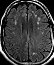
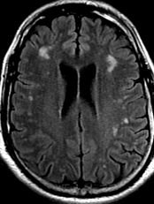
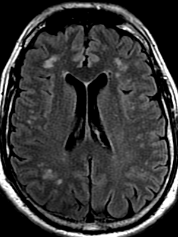
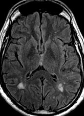
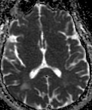
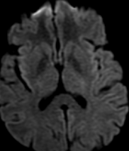
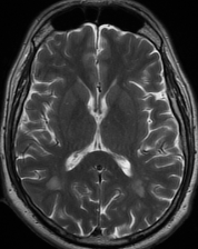
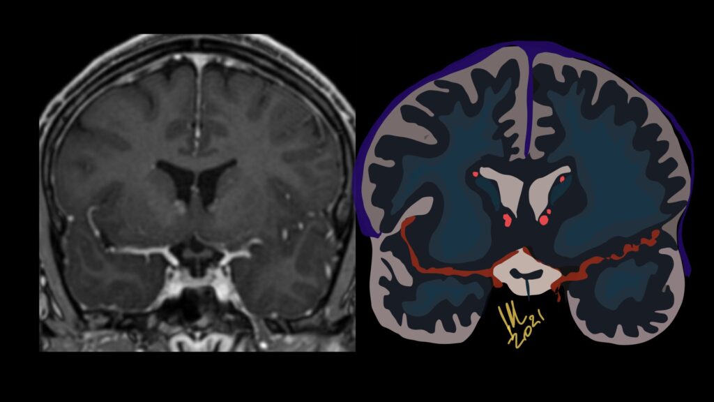
Encephalopathy and Cortical FLAIR signal abnormality- Covid-19

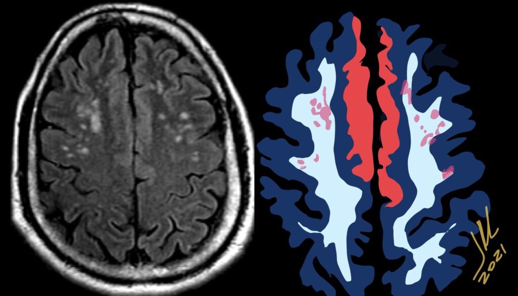
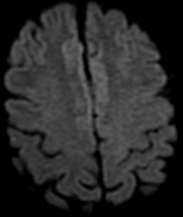
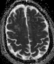
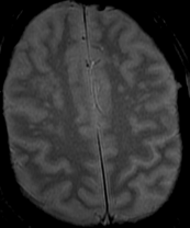
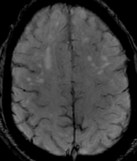
50M. patient who had covid course 70 days before without any respiratory manifestation only with persistent brain fog.

The most frequent diagnoses made at brain MR imaging in patients with COVID-19 are acute and subacute infarcts. Other common findings included a constellation of leukoencephalopathy and microhemorrhages, leptomeningeal contrast enhancement, and cortical FLAIR signal abnormality.

Meningitis and encephalitis are uncommon in patients with COVID-19 and
neurologic symptom. Also cortical FLAIR hyperintensity should be secondary to encephalitis. Nonspecific cortical pattern of T2 FLAIR hyperintense signal with associated restriction diffusion that may be caused by systemic toxemia, viremia and-or hypoxic effects.

Cortical FLAIR signal abnormality has a broad differential diagnosis and can include encephalitis, postictal state, PRES, as well as acute ischemia.

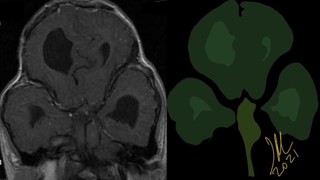
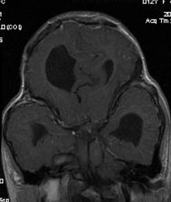
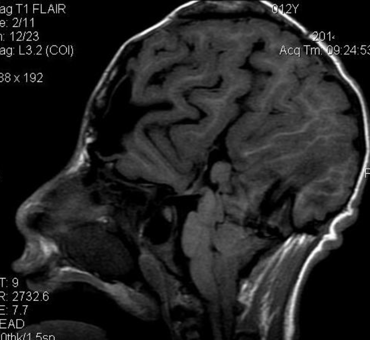
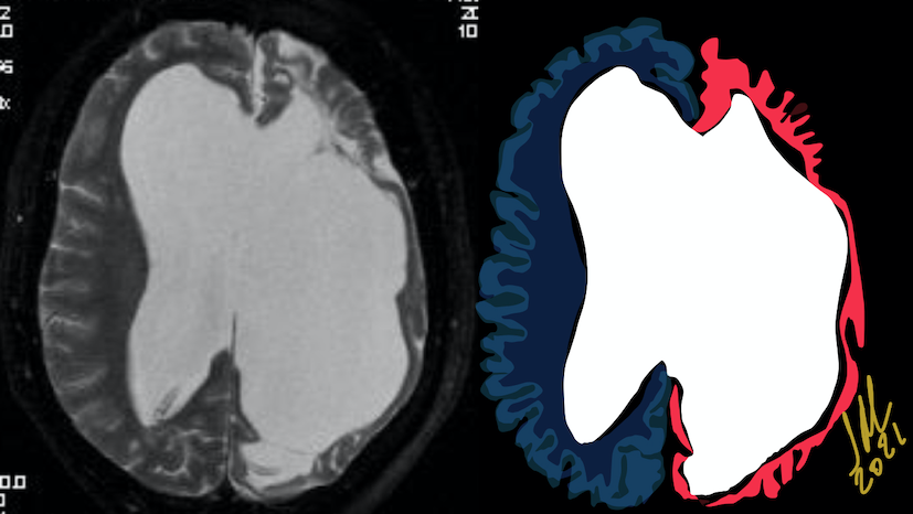
Pfeiffer syndrome type II

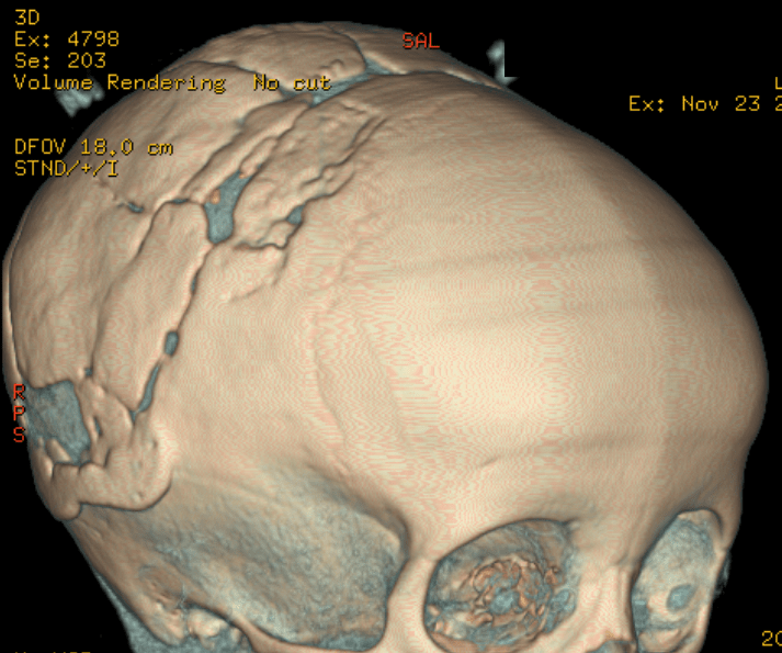
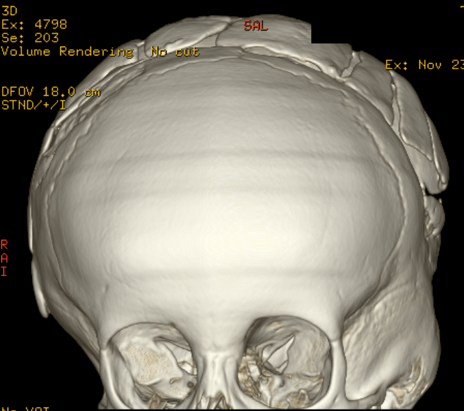
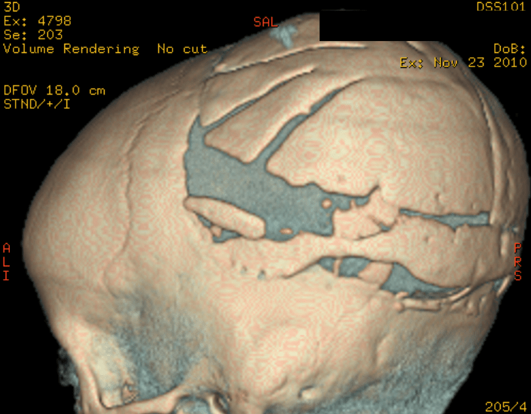
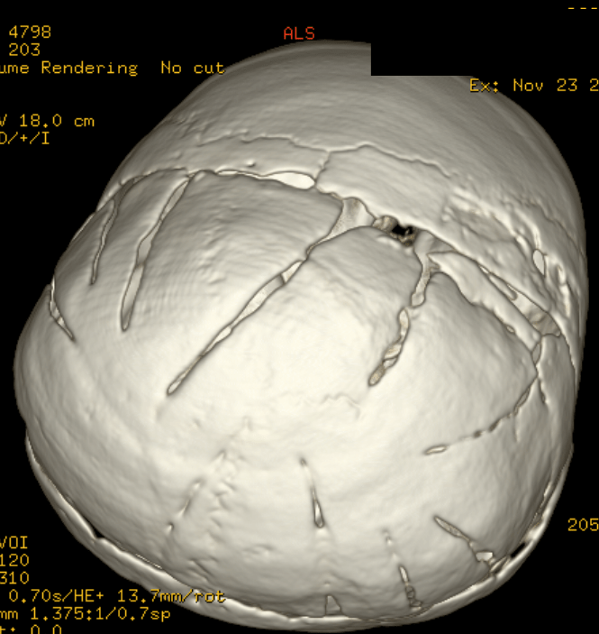
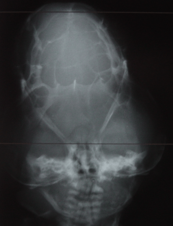
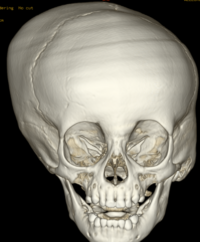
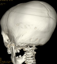
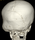
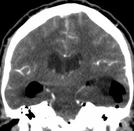
Pfeiffer syndrome is a form of pansynostosis of the cranial sutures with associated limb abnormalities. There are 3 types of Pfeiffer syndrome with type I (classic) being the mildest form of the disorder. Midface hypoplasia and abnormal skull shape are minimal in this form of the disease, and intelligence is more likely to be normal in these patients. Type II is more severe and results in the formation of a “cloverleaf” skull (our case), also known as kleeblattschädel, and the degree of cranial abnormality is more pronounced. Pfeiffer syndrome type II is commonly fatal in infancy due to its marked compression on the intracranial structures. Pfeiffer syndrome type III is also more severe than Pfeiffer syndrome type I; however, the cloverleaf skull phenotype is less common in Pfeiffer syndrome type III.

Pfeiffer syndrome type II is caused by a mutation in the gene for fibroblast growth factor receptor 2 (FGFR2) on the long arm of chromosome 10, a gene that encodes a cell membrane protein responsible for cell differentiation. Pfeiffer syndrome type I has an association with FGFR1, a different gene on the short arm of chromosome 8. Mutations in the FGFR genes are also implicated in Apert, Beare-Stevenson, Crouzon, Jackson-Weiss, and Muenke syndromes, all diseases that exhibit craniosynostosis and/or facial malformations to varying degrees. The pattern of inheritance for Pfeiffer syndrome is autosomal dominant with cases of Pfeiffer syndrome types II and III due primarily to sporadic de novo mutations.

The cloverleaf skull deformity (kleeblattschädel) was first described by Holtermuller and Wiedemann. The upper and lower leaves of the cloverleaf are formed by a ring of bone that divides the upper and lower leaves of the cloverleaf, and there is a honeycomb pattern of the inner vault of the skull. Crowding of the posterior fossa and subsequent hindbrain herniation are common. Kleeblattschädel can be seen in severe forms of Crouzon and Apert syndromes, Saethre-Chotzen syndrome, Carpenter syndrome, and thanatophoric dysplasia, though Pfeiffer syndrome has been reported to account for approximately 15%-20% of cases. Intellectual impairment observed in patients with cloverleaf skull is most likely a result of the intracranial mass effect from the calvarial deformity rather than an intrinsic brain abnormality, as normal intelligence by 4 years of age has been reported in some cases of patients undergoing a staged reconstruction of kleeblattschädel
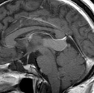
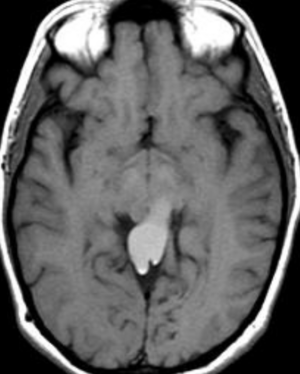
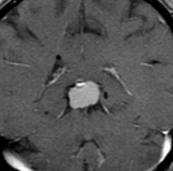
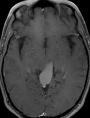
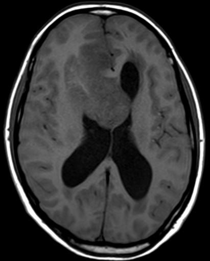
Pineal epidermoid cyst

The pineal localization is a very rare form of this intracranial lesion. It represents 0,2-1% of all intracranial tumors.  Cushing was the first to report the pineal localization of the epidermoid cyst in 1928. The clinical presentation is often characterized by parinaud’s syndrome and hydrocephalus. Hemiparesis and cerebellar signs can also be noticed

Craniosynostosis

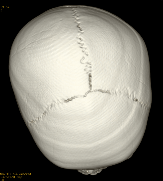
Craniosynostosis refers to the premature closure of the cranial sutures. The skull shape then undergoes characteristic changes depending on which sutures close early. 3D CT MIP can depict suture patency, extent of synostosis (ie, complete versus incomplete bone bridging), fracture extent and conspicuity, and three-dimensional calvarial deformity as a single set of projections in children with suspected craniosynostosis or skull fracture. 


Types of craniosynostosis

There are several types of craniosynostosis. Most involve the fusion of a single cranial suture. Some complex forms of craniosynostosis involve the fusion of multiple sutures. Most cases of multiple suture craniosynostosis are linked to genetic syndromes and are called syndromic craniosynostosis.

The term given to each type of craniosynostosis depends on what sutures are affected. Types of craniosynostosis include:

  • Sagittal (scaphocephaly). Premature fusion of the sagittal suture that runs from the front to the back at the top of the skull forces the head to grow long and narrow. Sagittal craniosynostosis results in a head shape called scaphocephaly and is the most common type of craniosynostosis.
  • Coronal. Premature fusion of one of the coronal sutures (unicoronal) that run from each ear to the top of the skull may cause the forehead to flatten on the affected side and bulge on the unaffected side. It also leads to turning of the nose and a raised eye socket on the affected side. When both coronal sutures fuse prematurely (bicoronal), the head has a short and wide appearance, often with the forehead tilted forward.
  • Metopic. The metopic suture runs from the top of the bridge of the nose up through the midline of the forehead to the anterior fontanel and the sagittal suture. Premature fusion gives the forehead a triangular appearance and widens the back part of the head. This is also called trigonocephaly.
  • Lambdoid. Lambdoid synostosis is a rare type of craniosynostosis that involves the lambdoid suture, which runs along the back of the head. It may cause one side of your baby’s head to appear flat, one ear to be higher than the other ear and tilting of the top of the head to one side.

Ref; Robertson RL, Ball Jr, WS, Barnes PD. Skull and brain. In: Kirks DR, Griscom NT, eds. Practical Pediatric Imaging: Diagnostic Radiology of Infants and Children, 3rd ed. Philadelphia: Lippincott-Raven; 1998:65-200

AJNR 2000, 21 (10) 1951-1954;

Tuberous sclerosis-subependymal nodules

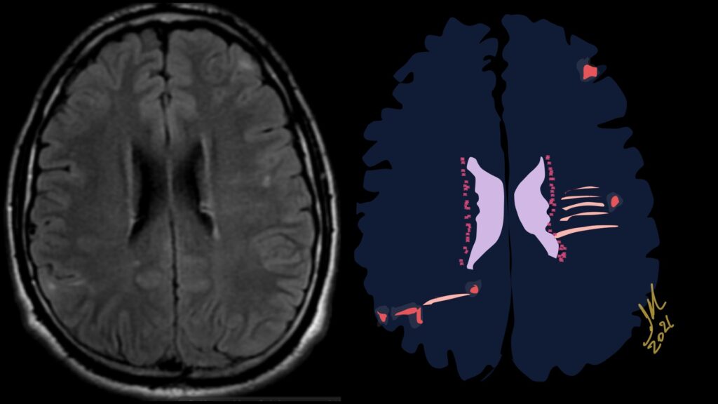
Imaging features, without any physical findings or symptoms, can also be used to make the diagnosis of TS. These unique diagnostic findings include the presence (on CT, MR, US) of multiple subependymal nodules (SENs; especially when calcified) or wedge-shaped cortical calcifications, intraventricular tumor consistent with subependymal giant cell astrocytoma (SGCA), and multiple cortical or subcortical “tubers” (especially if associated with subcortical white matter edema). In addition, typical linear abnormalities in the white matter may be found, Cystic lacune–like lesions have also been described in up to 44% of TS patients.

The SENs, although quite obvious on CT can be subtle on MR. The signal intensity patterns are variable and can change over time. Some SENs may have a “target” appearance on T2-weighted MRI, with a central hyperintensity reminiscent of the gyral core seen in the cortical tubers. the center of a SEN can be bright on the T1-weighted image.This appearance could be related to the paradoxical T1 shortening that occurs with dispersed microscopic calcifications. SENs show gadolinium enhancement, often clustered about the foramen of Monro. The SENs are histologically similar to the cortical hamartomas but are smaller and arise primarily in the striothalamate groove along the lateral margin of the lateral ventricles.

The SENs do not appear to grow; however, they do calcify progressively during the first two decades, so that by age 20 years virtually 100% are hyperdense, This presence of calcification allows SENs to be distinguished from the otherwise similar-appearing subependymal gray matter heterotopias. Heterotopic gray matter is isodense and isointense to normal gray matter, without contrast enhancement

Tuberous sclerosis complex (TSC) or Bourneville-Pringle syndrome is a Multisystem genetic disorder with epilepsy, multiorgan tumors, and hamartomas. Spectrum of central nervous system, hamartomas, all contain dysplastic neurons and giant (balloon) cells. Caused by mutation in TSC1 or TSC2 gene. Now considered infantile (developmental) tauopathy. Tau abnormally expressed in many of dysmorphic neurons and glial cells of TSC.

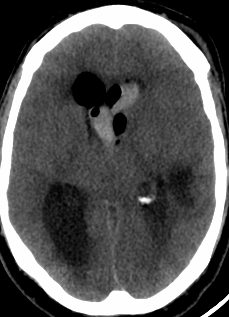
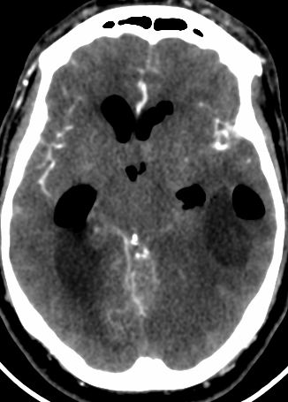
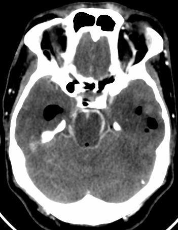
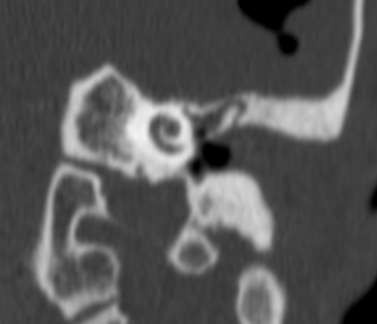
Otogenic brain abscess, hemorrhage and ventriculitis

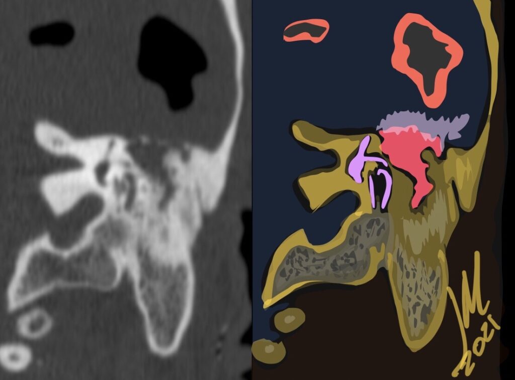
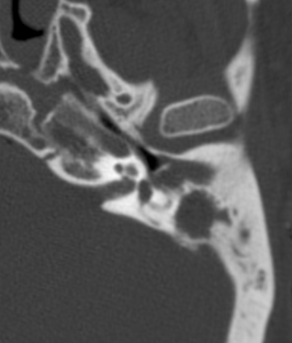
Otogenic brain abscess may occur as a complication of acute and chronic suppurative otitis media. Radiologists and Otolaryngologists should have a high index of suspicion for otogenic abscesses in patients with a history of chronic ear disease and new symptoms of fever, headache, and nausea.

Otitis media (OM) is a common otologic condition in pediatric and adult populations. Acute mastoiditis (AM) is a complication of otitis media in which infection in the middle ear cleft involves the mucoperiosteum and bony septa of the mastoid air cells. It can be divided into coalescent and non coalescent mastoiditis. In coalescent AM, infection causes osteolysis of the bony septa or cortical bone, which can further lead to intra and extracranial complications

The imaging technique of choice is CT for its sensitivity in detecting opacification and bone destruction. Its capability to differentiate among causes of opacification is poor. MR imaging provides additional imaging markers reflecting soft-tissue reaction to infection, major intramastoid signal changes, diffusion restriction or intramastoid, periosteal, or dural enhancement as well as brain abscess.

Complications of OM are classified as extracranial or intracranial. Brain abscess are commonly considered the second most common intracranial complication of OM after meningitis. Historically, it has been reported that 25% of brain abscesses in children were otogenic, whereas in adults it is thought that more than 50% of brain abscesses were otogenic.

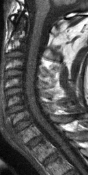
Lipoma of filum terminale

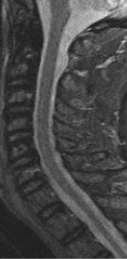
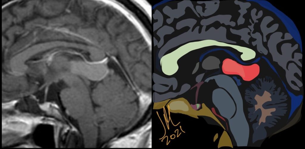
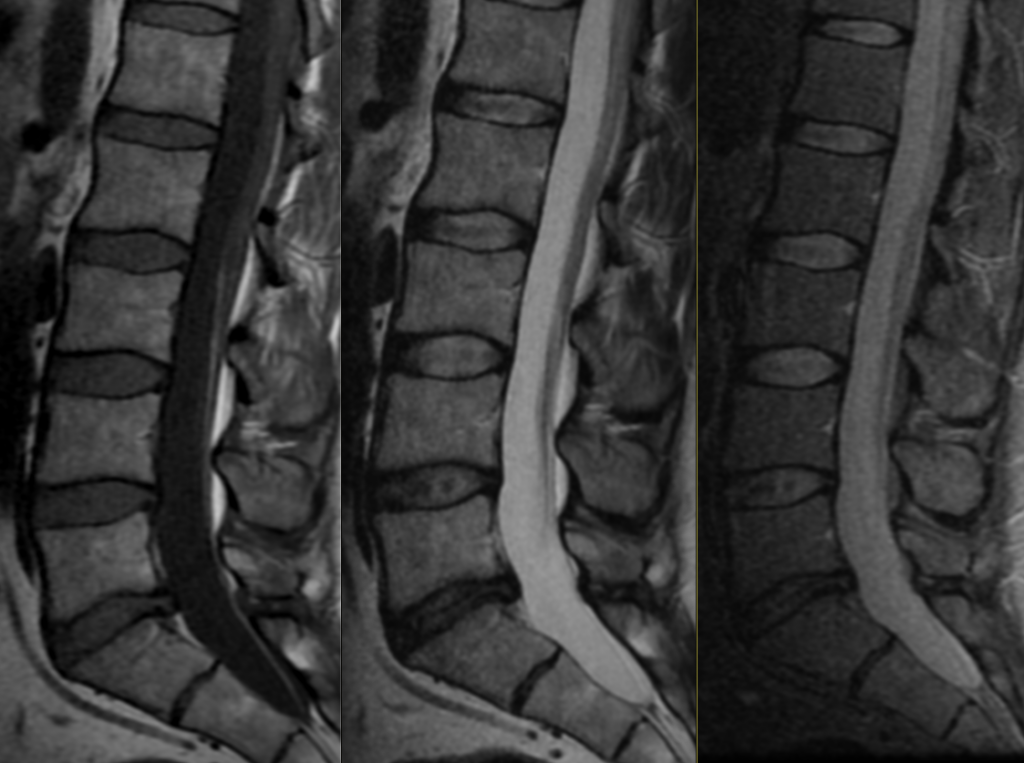
Filum terminale lipomas are increasingly being identified on imaging. The prevalence of isolated LFT is < 5%. There was no significant correlation between the thickness or length of LFT and the presence of neurological deficit. Most patients are asymptomatic and do not require frequent follow-up or surgical intervention

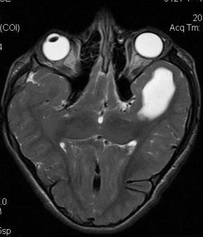
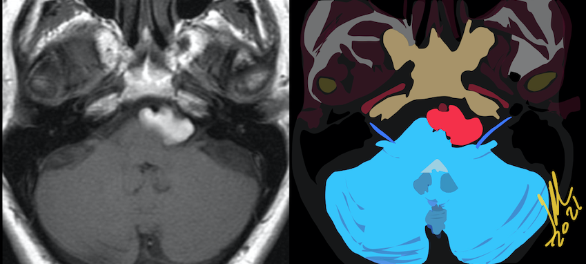
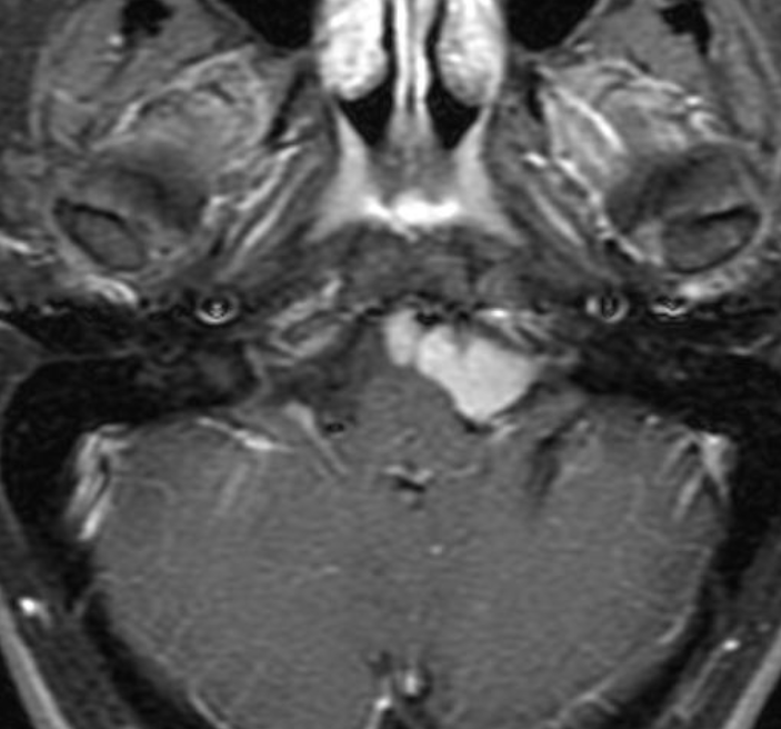
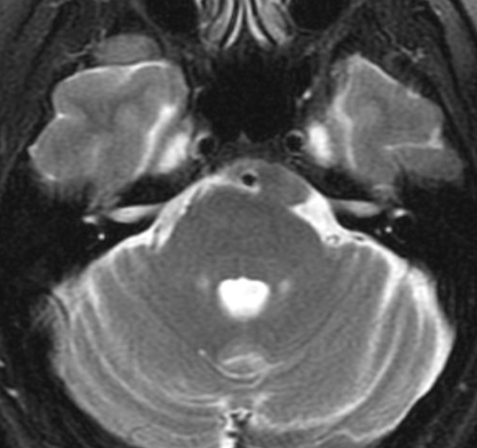
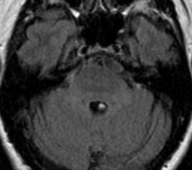
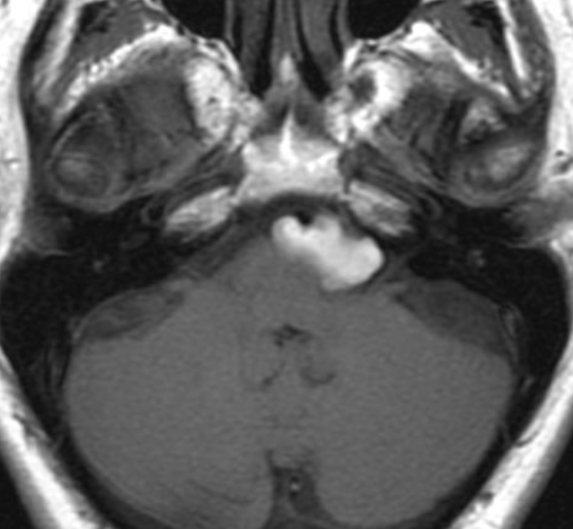
Hyperintense T1 Epidermoid Cyst in the prepontine area.

Epidermoid Cysts typically have long T1 (Hypo) and T2 (Hyper) relaxation times and have no enhancement on postcontrast MR images.

EC can show hyperintensity on T1 and T2 MR images, caused by a semiliquid cystic content with high protein concentration. It also can have high signal intensity on T1 and low signal intensity on T2 caused by a combination of high protein content and high viscosity.

EC can show low signal intensity on both T1- and T2-weighted images, with an usual composition of epithelial debris, crystals of cholesterol, and keratin

Dyke-Davidoff-Masson syndrome

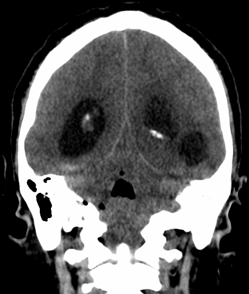
Dyke-Davidoff-Masson syndrome is a rare entity characterized by hemi cerebral atrophy/hypoplasia secondary to brain insult in fetal or early childhood period along with ipsilateral compensatory osseous hypertrophy and contralateral hemiparesis. The etiopathogenesis could be either vascular insult during intrauterine life resulting in hypoplasia of a cerebral hemisphere or acquired causes like trauma, infection, vascular abnormalities and intracranial hemorrhage in the perinatal period or shortly thereafter causing hemi cerebral atrophy

The diagnosis is based on the typical radiological features on CT and MRI scans which include cerebral hemiatrophy with dilated ipsilateral lateral ventricle. Also there is thickening of calvarium with enlargement of frontal, ethmoid and sphenoid sinuses and elevation of greater wing of sphenoid and petrous ridge.

The differential diagnoses are chronic Rasmussen encephalitis (chronic, progressive inflammation of brain of uncertain etiology) and sturge weber syndrome. However, Rasmussen encephalitis doesn’t show calvarial changes and sturge-weber syndrome additionally shows enhancing pial angiomas and cortical calcifications