Neurovascular Compression of Facial Nerve

Neurovascular compression syndrome (NVCS) is defined as a direct contact with mechanical irritation of cranial nerves by blood vessels.  By far the cases of neurovascular contact are clinically asymptomatic

NVCS can occur either at the root entry-exit zone 10%–90%,  adjacent cisternal segment 60%, Transitional zone 22%.

Microvascular decompression is highly effective in HFS and symptoms disappear after an operation in 90%–95% of cases; however, recurrence is seen in up to 25% of patients.
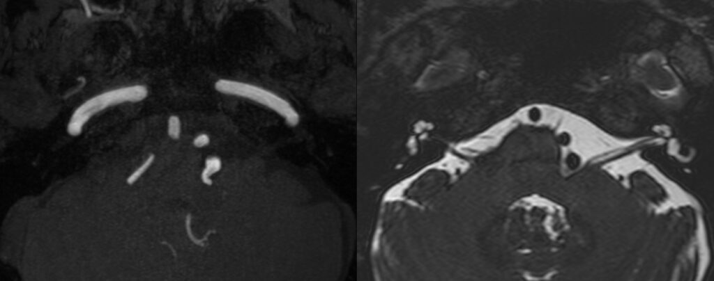
high-resolution 3D T2-weighted imaging with 3D time-of-flight angiography and 3D T1-weighted gadolinium-enhanced sequences is considered the standard of reference for the detection of neurovascular compression 

The transition zone between central and peripheral myelin is an anatomic area with increased mechanical vulnerability, which is of particular interest in the context of symptomatic NVCS. The transitional zone look to be the more relevant and vulnerable anatomic structure, and it is not always located in the same position as the root entrance-exit zone (TZ). Exact anatomic knowledge of the position and morphology of the TZ is of fundamental importance for the interpretation of neuroimaging findings in suspected NVCS. TZ of VII CN to be about <1mm to REZ.

To remember TZ location from REZ:

CN V = 2mm. CN VIII = 1mm. CN VIII= 10mm. CN IX= 15mm
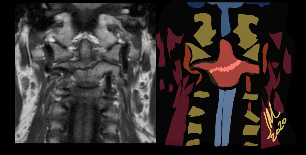
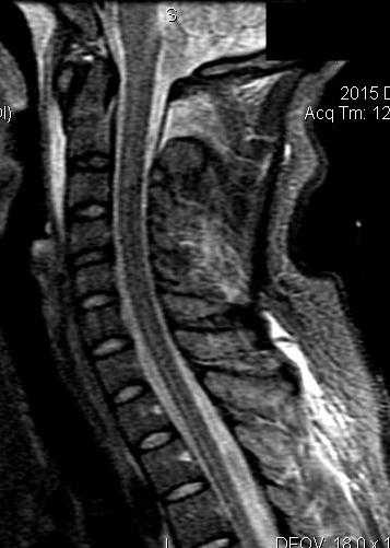
STIR Sequence for Acute Type II-III

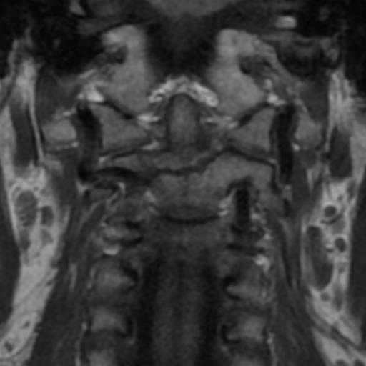
Type III

Because MR imaging and the STIR sequence are used to determine fracture acuity and, therefore, treatment recommendations, it is important that we understand the limitations of this sequence.The STIR sequence is relied on to reveal marrow edema related to an acute fracture. In addition to the increased detection of soft-tissue injury, the STIR sequence is exquisitely sensitive to bone marrow pathology. This feature may be useful in identifying the acuity of a fracture. The STIR sequence, while sensitive for the detection of acute type II-III odontoid fracture in patients younger than 50 years, is significantly less sensitive in older patients, particularly those with osteopenia. In older patients The combination of the vascular watershed and the lack of a periosteal blood supply contributes to overall poor vascularity in the odontoid, which contributes to the absence of bone marrow edema and, subsequently, the absence of STIR hyperintensity in the setting of acute fracture in the older population. Evaluating all the sequences is imperative and the combination of the t1 sequence in the coronal and sagittal plane with the stir sagittal and coronal sequence is very useful.

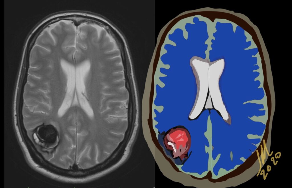
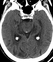
Atypical hemorrhage due to Zabramski type V Cavernoma

Type V indicate parts of the actual CCMs that are visible in the center of the hemorrhage; however, the CCM is not fully distinguishable from hemorrhage. Type 5 has a high hemorrhage risk of 24%

Hemorrhagic events particularly those found in relatively young patients ought to be characterized further, and cavernous angioma should be considered among the possible etiologies. Also because of their propensity to act as an epileptogenic focus CMs should always be considered in the workup of a patient with a seizure disorder, especially if the patient is aged 20-40 years.

If a recent bleed has occurred then surrounding edema or mass effect may be present. The lesions show a variable pattern of enhancement. Contrast-enhanced images help delineate any potential associated developmental venous anomalies. This is critical for preoperative surgical planning as the un-deliberate resection of DVAs may compromise normal cortical venous drainage and thus lead to brain venous infarction. Angiographically these lesions are occult and usually show nonspecific findings like capillary blush or early draining vein

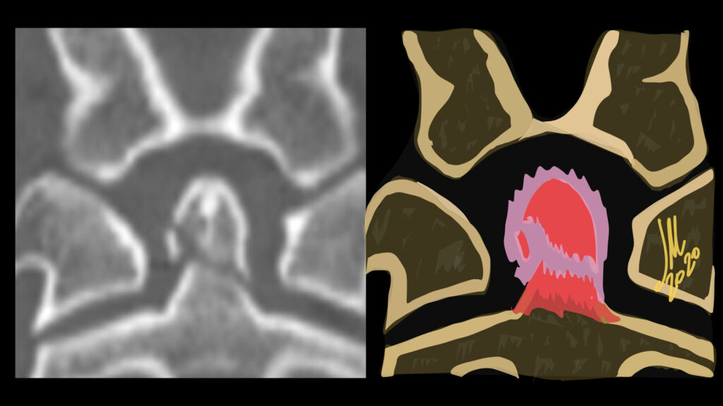
Odontoid type 2 fracture

Fracture through the base of the dens. Type II odontoid fractures are potentially unstable with subsequent myelopathy and spinal injury risk. Higher risk of nonunion with comminuted fracture, age > 50 or more than 5 mm displacement.
Odontoid fracture is a very common cervical injury, accounting approximately for 9%–15% of all cervical fractures. These injuries result from an hyperextension or hyperflexion mechanism of the cervical spine in low-energy impacts such as falls for elderly patients or in high-energy impacts such as motor vehicles accidents for young people.

The classification scheme of odontoid fractures described by Anderson and D’Alonzo is the one most commonly used. However there is a new classification by Grauer who proposed a better difference between type 2 -3.

Cervical spine trauma is most commonly evaluated with CT with MPR reconstructions 1-3mm.

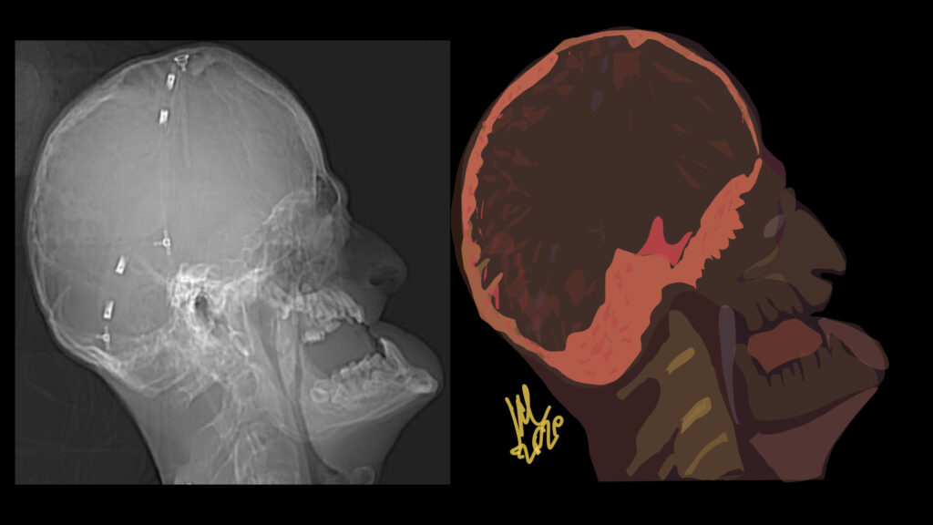
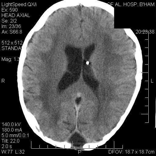
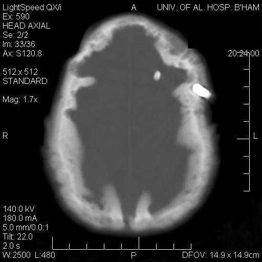
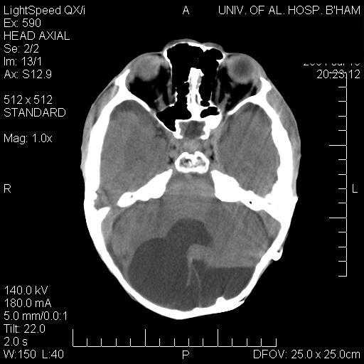
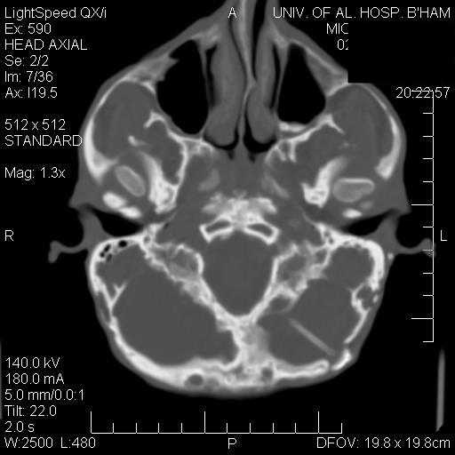
Hutchinson-Gilford Progeria Syndrome

Hutchinson-Gilford Progeria Syndrome (HGPS) is a rare syndrome of segmental premature aging. Children appear healthy at birth and within the first year of life develop severe failure to thrive with sclerodermatous skin changes. Classic craniofacial features develop with age and underlying disease culminates in death from myocardial infarction or stroke.

Thinning and mottling of the calvarium, prominent vascular markings, delayed closure of the anterior and posterior fontanels, widening of calvarial sutures, and a J-shaped sella, optic nerve kinking and hypotelorism, short mandibular rami in combination with flattened mandibular condyles are observed.

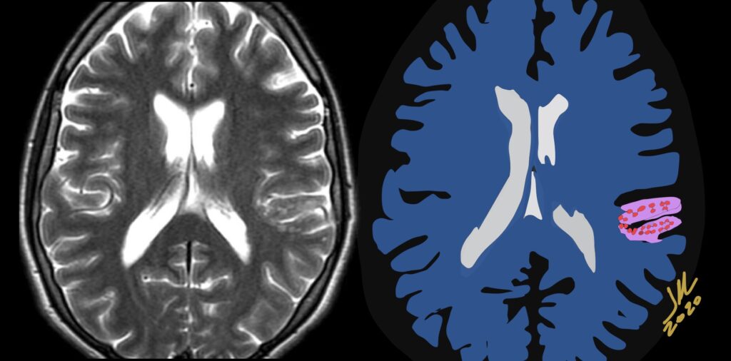
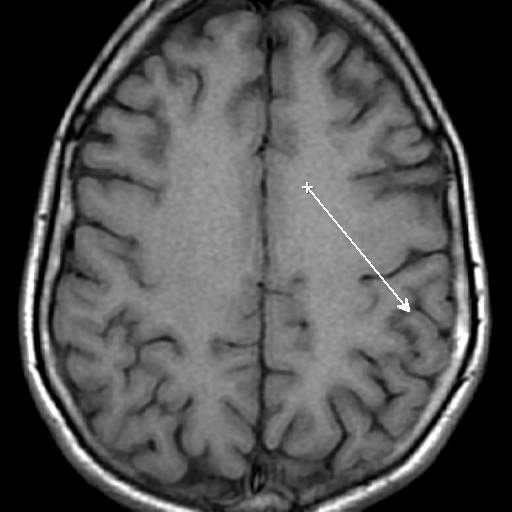
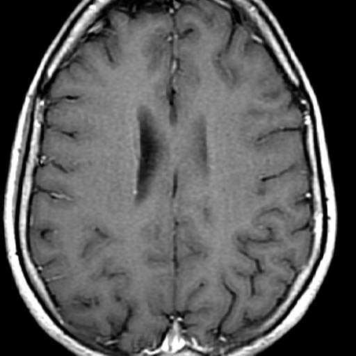
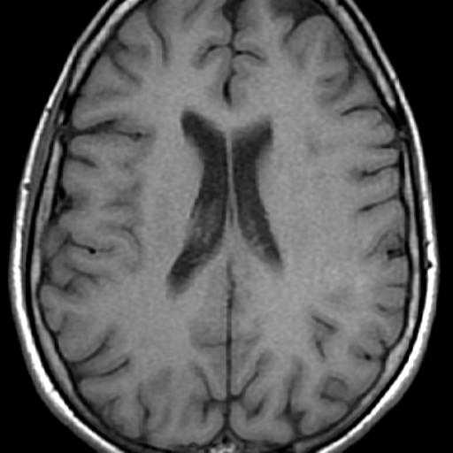
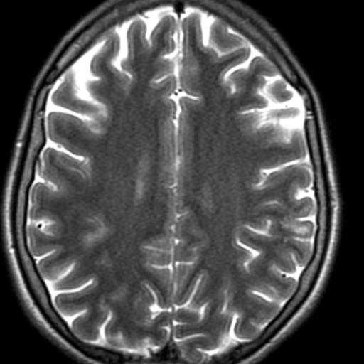
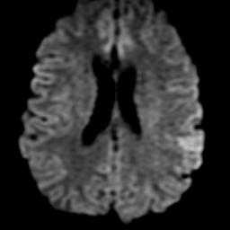
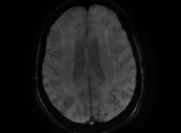
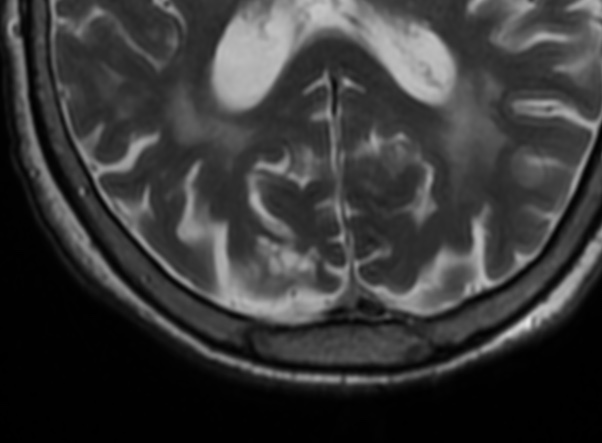
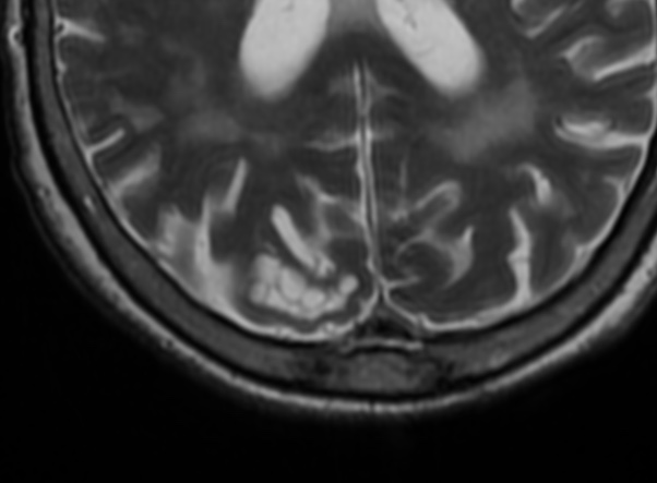
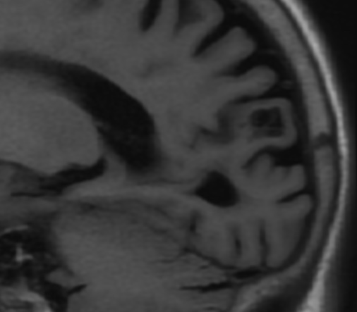
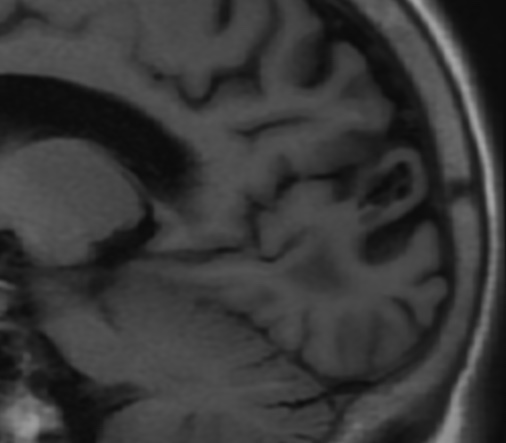
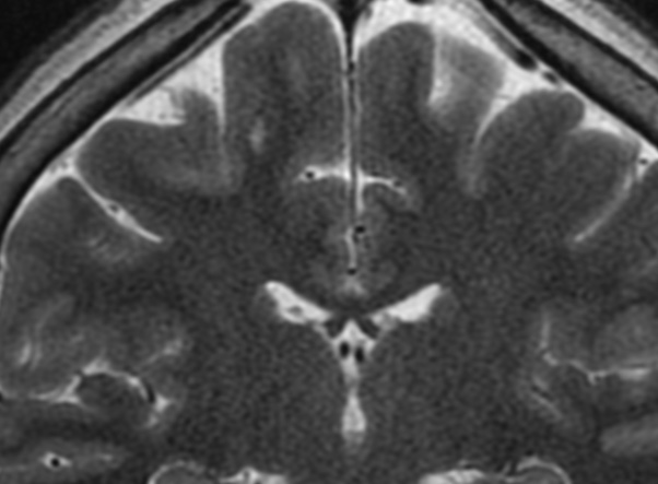
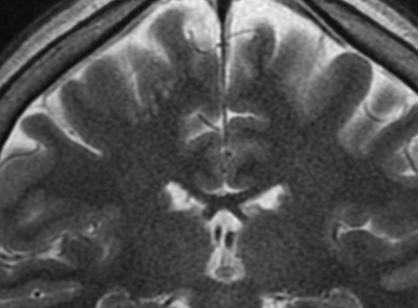
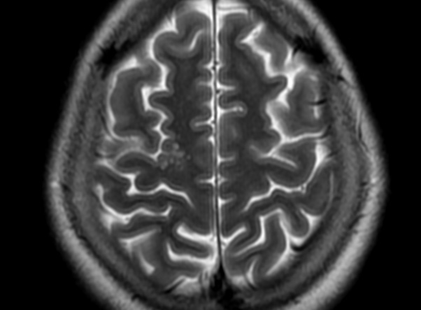
Multinodular and vacuolating neuronal tumors

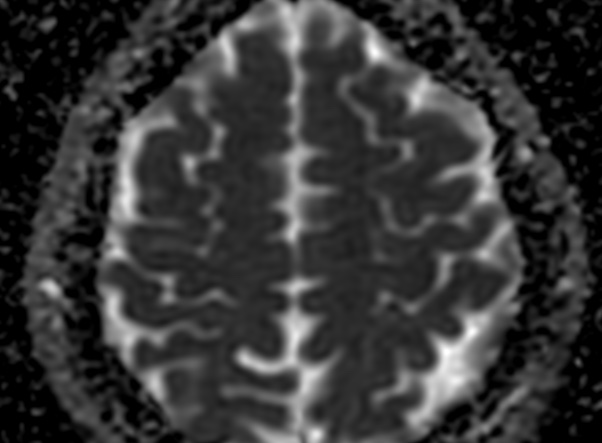
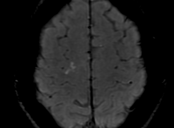
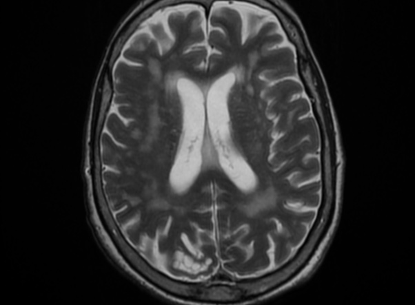
Multinodular and vacuolating neuronal tumors of the cerebrum (MVNT) are superficial neuronal tumors in adults that were first documented in 2013. It is characterized by a group of intra-axial nodules with mixed neuro-glial cells demonstrating internal vacuolating morphology. The lesions are benign and non progressive. the nodules are hypointense on T1, hyperintense on T2, and are not suppress on FLAIR. There are no enhancement, susceptibility, or DWI restriction. There are no edema or mass effect.