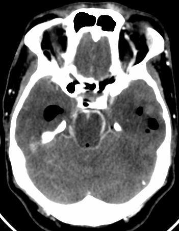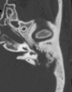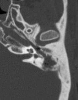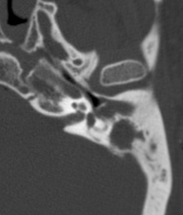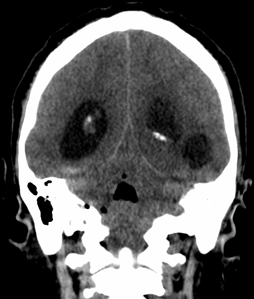
Otogenic brain abscess, hemorrhage and ventriculitis

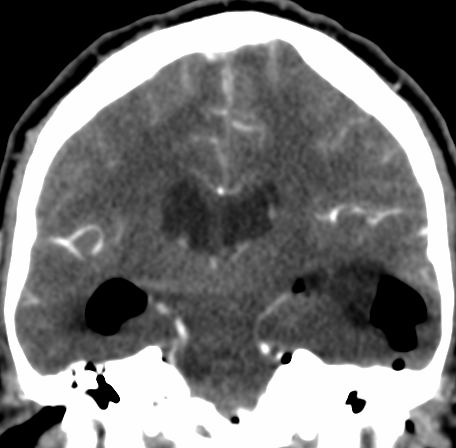
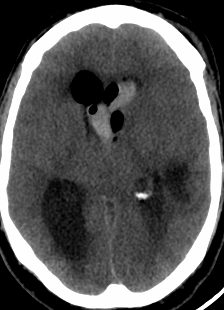
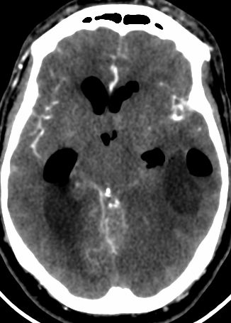
Otogenic brain abscess may occur as a complication of acute and chronic suppurative otitis media. Radiologists and Otolaryngologists should have a high index of suspicion for otogenic abscesses in patients with a history of chronic ear disease and new symptoms of fever, headache, and nausea.

Otitis media (OM) is a common otologic condition in pediatric and adult populations. Acute mastoiditis (AM) is a complication of otitis media in which infection in the middle ear cleft involves the mucoperiosteum and bony septa of the mastoid air cells. It can be divided into coalescent and non coalescent mastoiditis. In coalescent AM, infection causes osteolysis of the bony septa or cortical bone, which can further lead to intra and extracranial complications

The imaging technique of choice is CT for its sensitivity in detecting opacification and bone destruction. Its capability to differentiate among causes of opacification is poor. MR imaging provides additional imaging markers reflecting soft-tissue reaction to infection, major intramastoid signal changes, diffusion restriction or intramastoid, periosteal, or dural enhancement as well as brain abscess.

Complications of OM are classified as extracranial or intracranial. Brain abscess are commonly considered the second most common intracranial complication of OM after meningitis. Historically, it has been reported that 25% of brain abscesses in children were otogenic, whereas in adults it is thought that more than 50% of brain abscesses were otogenic.
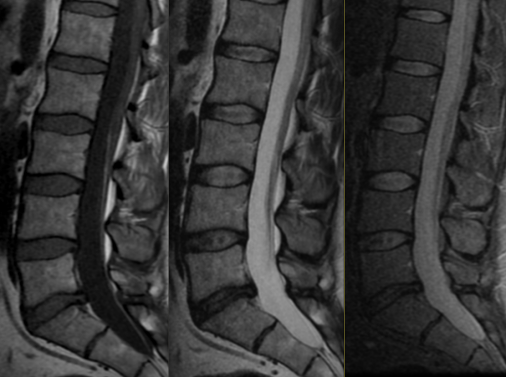
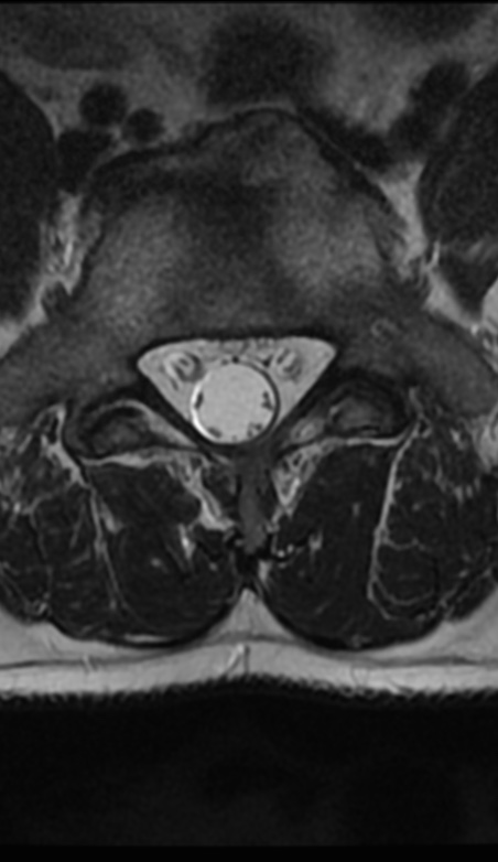
Lipoma of filum terminale

Filum terminale lipomas are increasingly being identified on imaging. The prevalence of isolated LFT is < 5%. There was no significant correlation between the thickness or length of LFT and the presence of neurological deficit. Most patients are asymptomatic and do not require frequent follow-up or surgical intervention

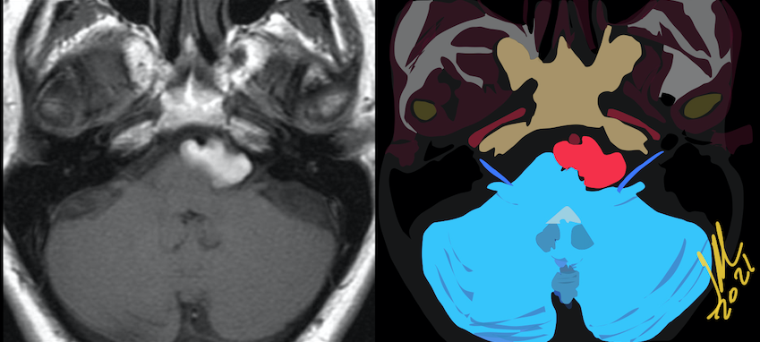
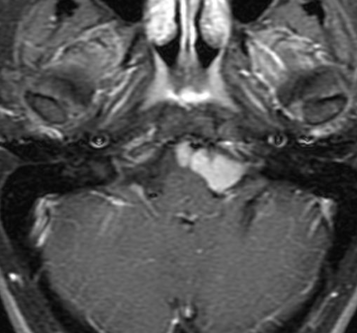
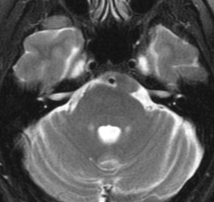
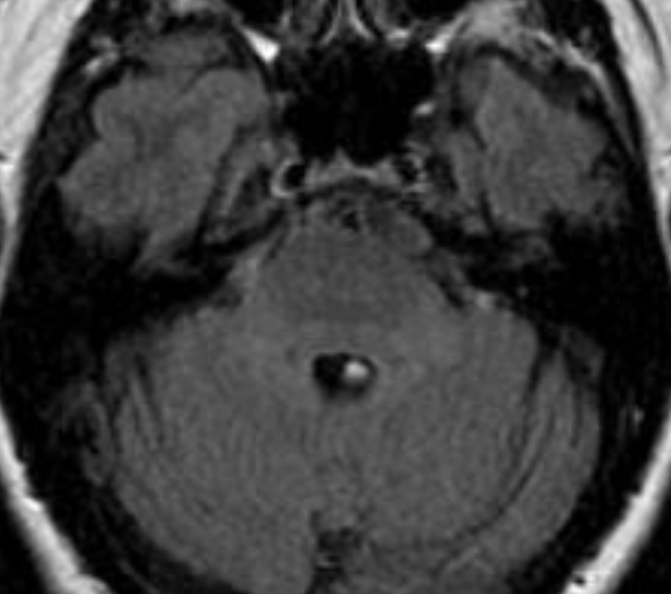
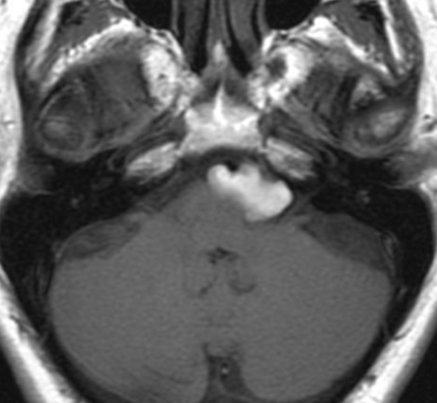
Hyperintense T1 Epidermoid Cyst in the prepontine area.

Epidermoid Cysts typically have long T1 (Hypo) and T2 (Hyper) relaxation times and have no enhancement on postcontrast MR images.

EC can show hyperintensity on T1 and T2 MR images, caused by a semiliquid cystic content with high protein concentration. It also can have high signal intensity on T1 and low signal intensity on T2 caused by a combination of high protein content and high viscosity.

EC can show low signal intensity on both T1- and T2-weighted images, with an usual composition of epithelial debris, crystals of cholesterol, and keratin

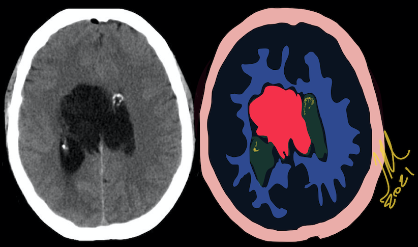
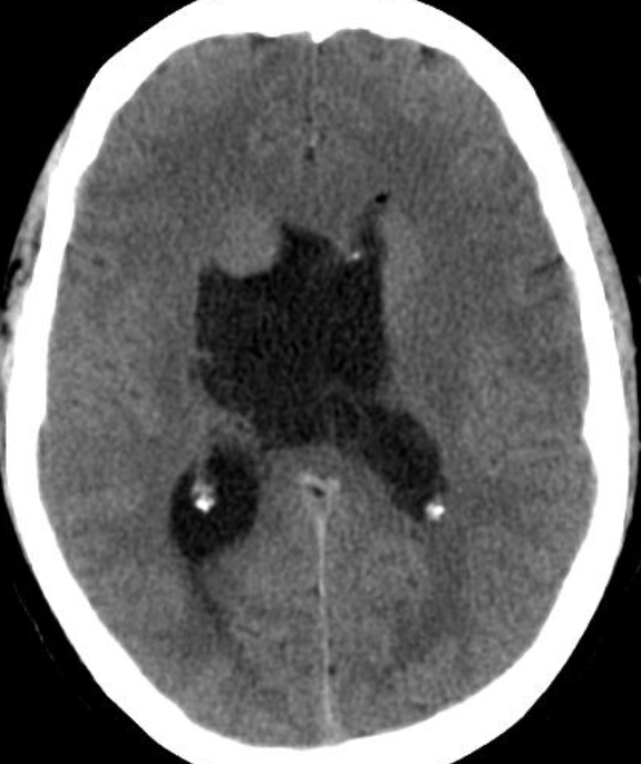
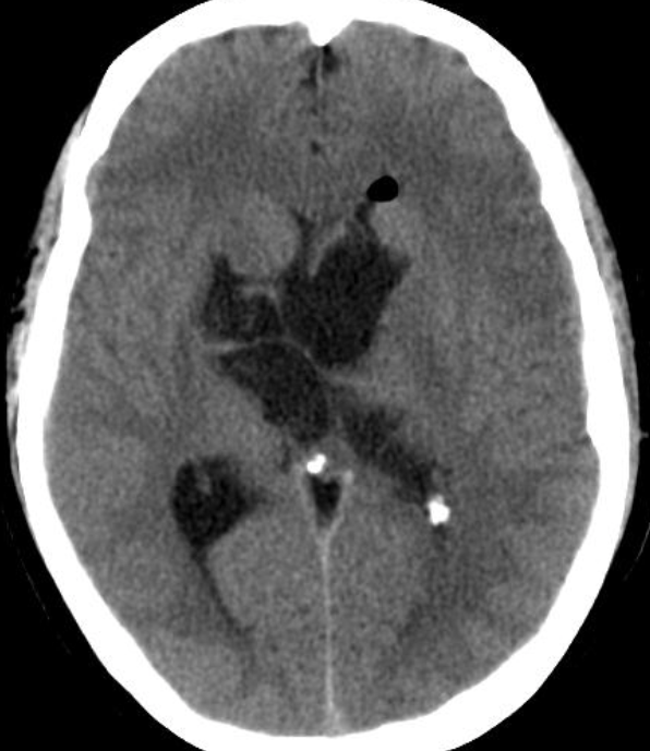
Intraventricular Epidermoid Cyst

Epidermoid cysts, are rare benign tumors developed from ectodermic inclusions. They usually sit at the ponto cerebellar angle, the para-sellar region and the temporal fossa. Their location at the intraventricular is very rare.  Most of the lateral intraventricular epidermoids are silent and/or do not produce hydrocephalus.

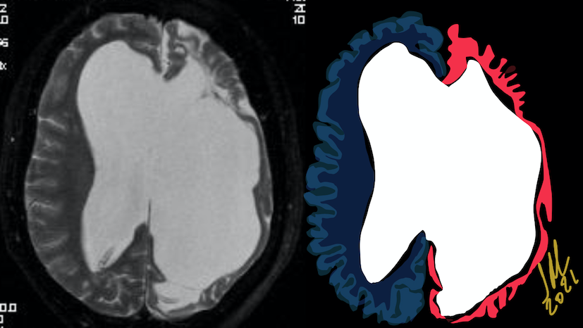
Dyke-Davidoff-Masson syndrome

Dyke-Davidoff-Masson syndrome is a rare entity characterized by hemi cerebral atrophy/hypoplasia secondary to brain insult in fetal or early childhood period along with ipsilateral compensatory osseous hypertrophy and contralateral hemiparesis. The etiopathogenesis could be either vascular insult during intrauterine life resulting in hypoplasia of a cerebral hemisphere or acquired causes like trauma, infection, vascular abnormalities and intracranial hemorrhage in the perinatal period or shortly thereafter causing hemi cerebral atrophy

The diagnosis is based on the typical radiological features on CT and MRI scans which include cerebral hemiatrophy with dilated ipsilateral lateral ventricle. Also there is thickening of calvarium with enlargement of frontal, ethmoid and sphenoid sinuses and elevation of greater wing of sphenoid and petrous ridge.

The differential diagnoses are chronic Rasmussen encephalitis (chronic, progressive inflammation of brain of uncertain etiology) and sturge weber syndrome. However, Rasmussen encephalitis doesn’t show calvarial changes and sturge-weber syndrome additionally shows enhancing pial angiomas and cortical calcifications

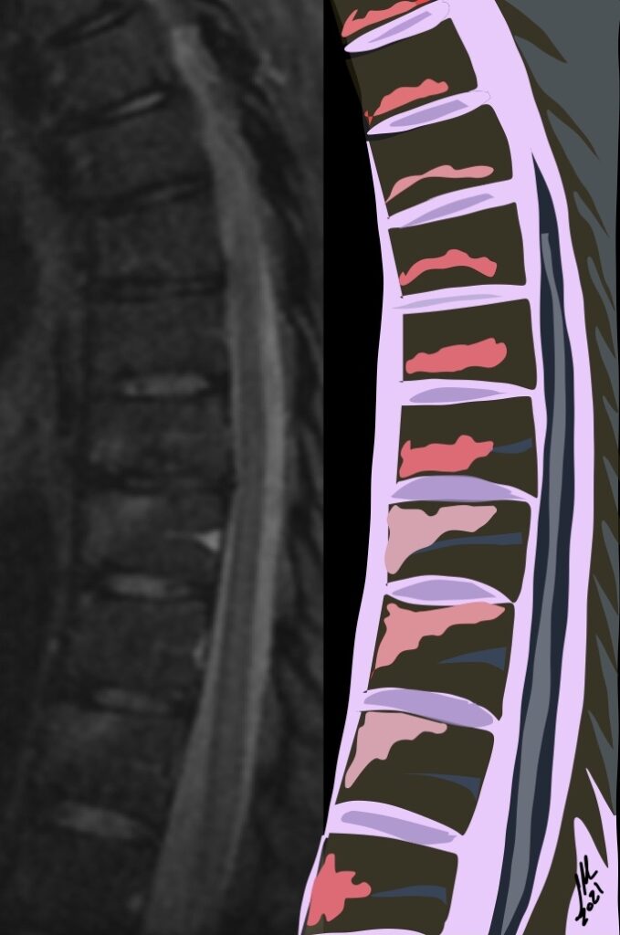
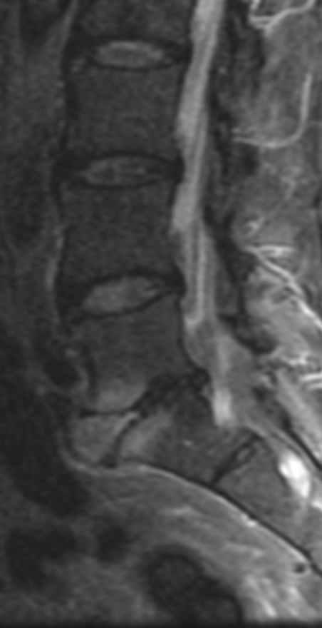
Andersson lesions

AL are characterized by high signal intensity at the cortical plates adjacent to intervertebral disks on STIR images and T1+C FAT SAT. High signal intensity may also be visible in the center or throughout the intervertebral space, simulating inflammatory diskitis. Andersson lesions are observed in 33% of patients with spondyloarthritis, and this finding has a specificity of 59% for this group of diseases


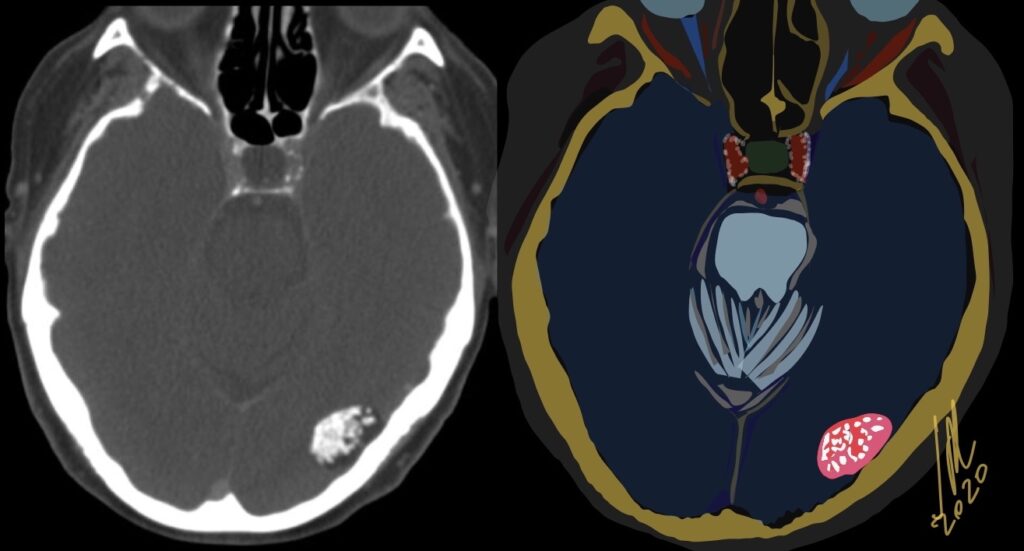
Atypical Epidermoid cyst

Epidermoid cysts have a thin squamous lining, which only rarely contains calcifications. These cysts contain debris from the desquamation of their squamous epithelial lining. The debris consists of mostly keratin, a proteinaceous material, and some cholesterol, a lipid material derived from the breakdown of cell membranes. On CT, the epidermoid cysts appeared hypodense with no evidence of enhancement after contrast material and edema is absent. 1.5% located in Cerebral hemispheres and 25% are calcified.

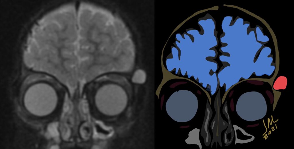
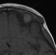
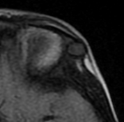
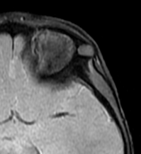
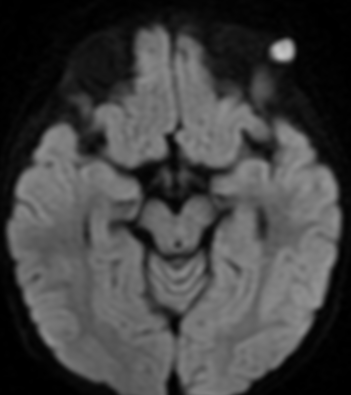
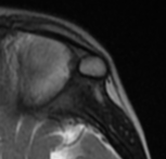
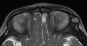
Intradiploic Epidermoid Inclusion Cyst

10% of intracranial epidermoids are extradural, being encountered most often in the skull as intradiploic masses in the temporal, occipital, parietal and frontal bones. Most manifest as painless, visible masses and are usually found along the lines of embryologic fusion; the zygomaticofrontal and the frontoethmoidal sutures

On MRI, epidermoids are hypointense on T1 hyperintense on T2 and maybe the most important thing have high signal intensity on DWI.

Differential diagnoses include dermoid cysts, eosinophilic granulomas, cholesterol granulomas, hemangiomas, aneurysmal bone cysts, fibrous dysplasia and eosinophilic granuloma.

Anterior lumbar disc herniation

Could be important to consider the possibility of discogenic visceral pain secondary to anterior herniation of the lumbar disc when forming a differential diagnosis for seemingly idiopathic abdominal pain. Visceral pain is a very frequent cause for medical consulting. Afferent fibers innervating viscera project to the central nervous system via sympathetic nerves. The lumbar sympathetic nerve trunk lies in front of the lumbar spine. Thus, it is possible for patients to suffer visceral pain originating from sympathetic nerve irritation induced by anterior herniation of the lumbar disc.
The most important thing is that it is always a diagnosis of exclusion and highly questioned.

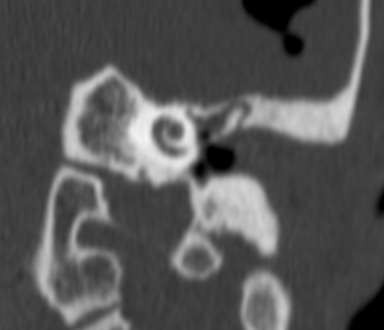
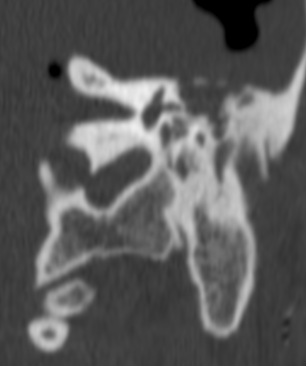
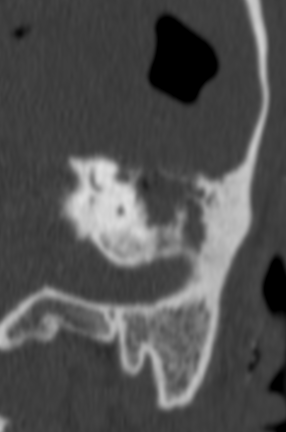
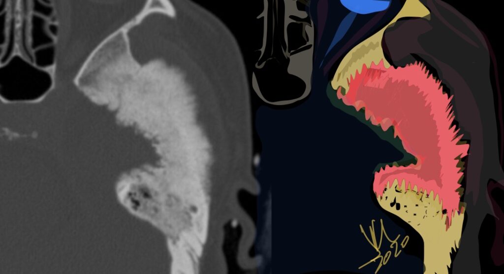
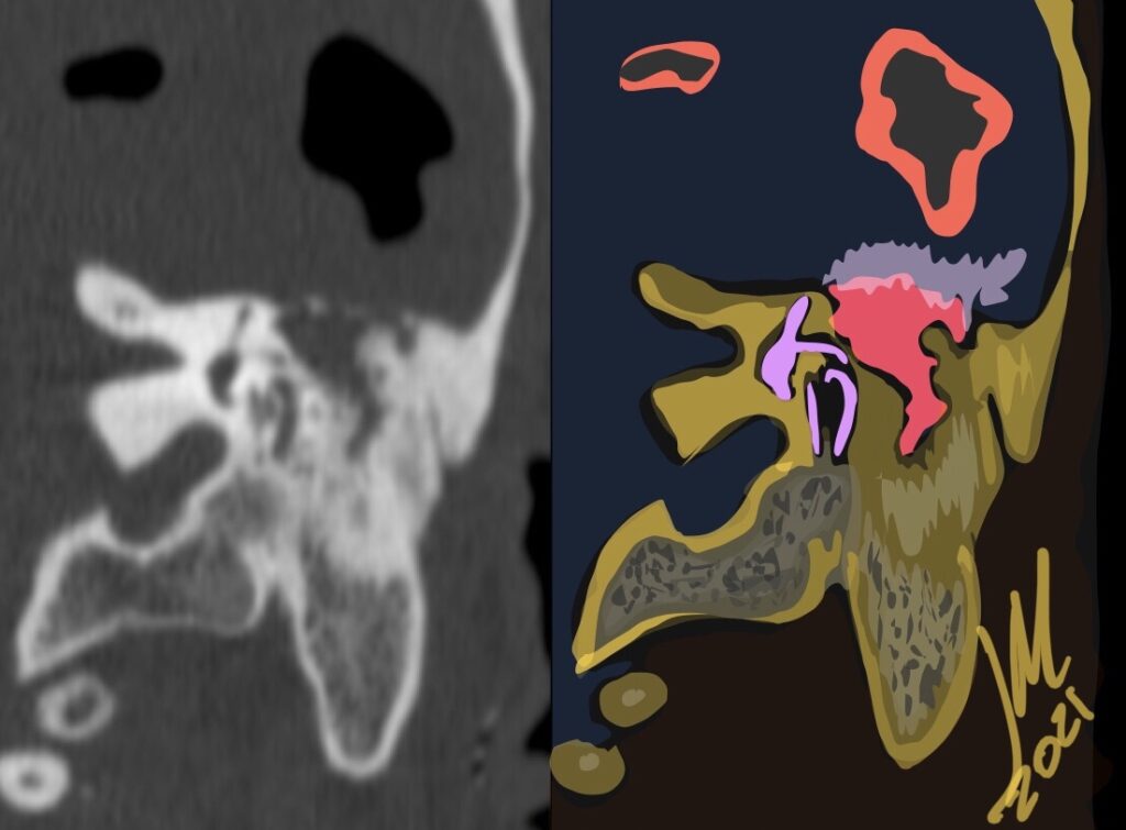
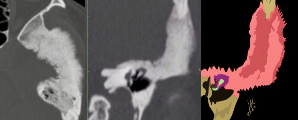
Tegmen Tympani Meningioma-CT

Meningioma primary to the tegmen tympani arise from the floor of the middle cranial fossa and spread inferomedially into the middle ear cavity.

CT features included thickening of the tegmen tympani, The internal trabecular architecture of the involved bone is preserved (trabecular hyperostosis). The inner margin of the involved calvaria along the lateral aspect of the middle cranial fossa is irregular. A middle ear cavity soft-tissue mass resulted in ossicular encasement. There is no ossicular erosion or destruction. Facial nerve canal encroachment is present.

Tegmen tympani meningioma bone changes on CT can be confused with fibrous dysplasia of this area. Preservation of internal trabecular architecture distinguishes it from the ground-glass attenuation seen in typical fibrous dysplasia.

 The other dominant consideration for tegmen tympani meningioma on CT is cholesteatoma; however, on CT, meningiomas lack the bone and ossicular destructive changes. These all lack the characteristic tegmen tympani thickening on CT and dural enhancement along the floor of the middle cranial fossa seen with meningioma on enhanced MR imaging. Enlargement of the facial canal typical of schwannoma is not seen with meningioma. Facial nerve hemangioma causes amorphous honeycomb bone changes on CT in larger lesions, which differ from the trabecular hyperostosis of tegmen tympani meningioma. Ossifying variants of hemangioma may have spicules of lamellar bone, which could more easily mimic meningioma on CT; however, most facial nerve hemangiomas are primarily centered in the geniculate fossa. This location should help distinguish them from meningioma