Giant Cavernous Malformation 

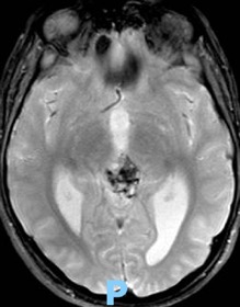
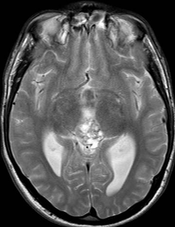
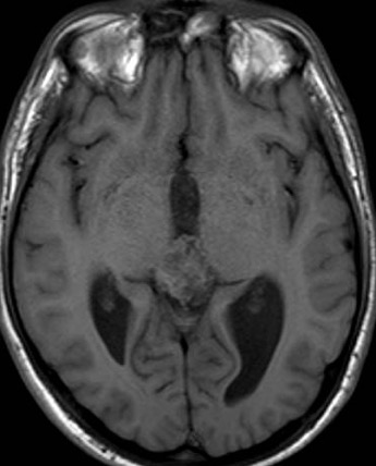
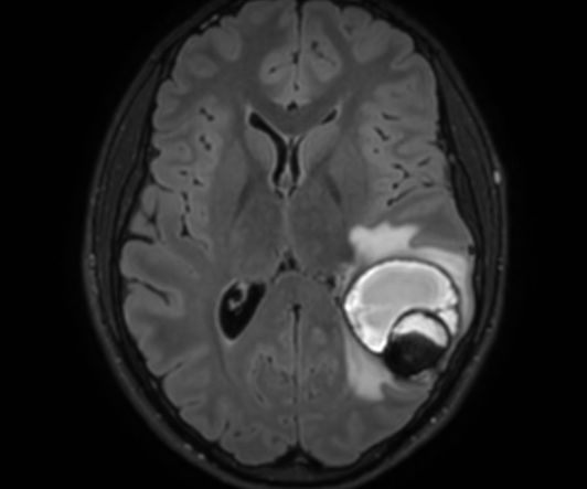
The cavernous malformation (CM), also known as cavernous angioma or cavernoma, is a vascular malformation characterized by the presence of sinusoid-like capillary vessel containing blood in very sluggish circulation.

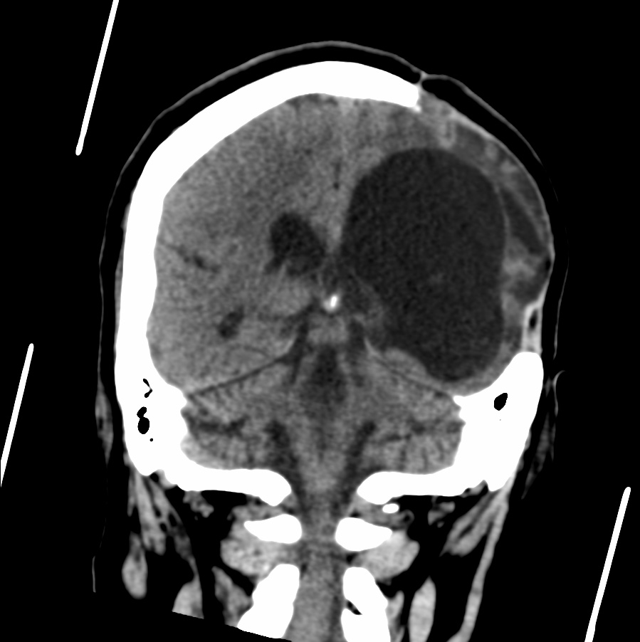
CMs vary in size from a few millimeters to a few centimeters. Few data can be found about the size of these malformations. Kim et al. reported the size of cavernoma between 1mm and 75 mm, with a mean size 14.2 mm. The majority of cavernous malformations are small but it may reach significant size. Unlike giant aneurysms, defined as having a diameter of at least 25 mm, no threshold dimension has been accepted for giant cavernous malformation (GCM).

Lawton et al., although arbitrary, defined a GCM as a cavernoma with a diameter greater than 6 cm. GCMs are very rare and usually not considered in the differential diagnosis of large tumor.

Cerebral CMs rarely attain large dimension. The mechanism by which they enlarge is probably recurrent bleeding, followed by organization of the clot, pseudocapsule formation, and secondary expansion. However, it was also reported that CMs can show expansile growth without any evidence of a hemorrhagic event and mimic neoplasm.

Reference:
10.3340/jkns.2008.43.4.198

Posttraumatic CSF leaks

CSF leaks can occur in patients of all ages. Most patients reveal a history of trauma or surgical procedures. However, spontaneous CSF leaks are not uncommon and are often described in middle-aged obese women with signs of intracranial hypertension.

Cerebrospinal fluid (CSF) leakage occurs when there is a tear or hole in the membranes that surround the brain and spinal cord, which causes the fluid to leak out. Traumatic CSF leaks can occur as a result of head or spinal injuries.

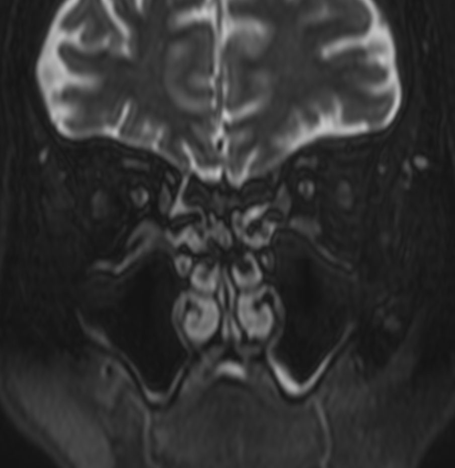
MRI (Magnetic Resonance Imaging) can be used to detect CSF leaks and identify the location of the tear or hole. The following are some of the common MRI findings associated with CSF traumatic leaks:

  1. Diffuse meningeal enhancement: This refers to the enhancement of the meninges, the membranes that surround the brain and spinal cord, which can be seen on contrast-enhanced MRI scans.
  2. Disruption of the dural sac: The dural sac is a tough, fibrous membrane that encloses the spinal cord. MRI may reveal a defect or disruption in the dural sac, which can cause CSF to leak out.
  3. Intracranial air: If there is a fracture in the skull base, air may enter the cranial cavity, which can be seen on MRI as hypointense areas.
  4. Distended pituitary gland: In some cases, a CSF leak can cause the pituitary gland to become enlarged or distended, which can be seen on MRI.

It is important to note that MRI findings alone may not be sufficient to diagnose a CSF leak. A combination of clinical symptoms, physical examination, and imaging tests may be necessary to make an accurate diagnosis.

Various imaging tests are used for identification of CSF leak. These include radionuclide cisternography, HRCT of the skull base, CT cisternography, and MR cisternography. 

Reference:
10.4103/ijri.IJRI_380_16

Leigh syndrome

Leigh syndrome is a progressive neurodegenerative disorder of childhood with an estimated incidence of 1:40,000 births.

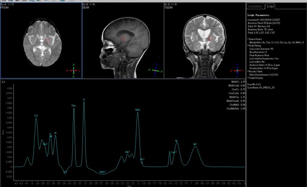
Leigh syndrome is the most common clinical phenotype of mitochondrial disorders in childhood. The diagnostic criteria are (1) progressive neurological disease with motor and intellectual developmental delay; (2) signs and symptoms of brainstem and/or basal ganglia disease; (3) raised lactate levels in blood and/or cerebrospinal fluid (CSF); and (4) characteristic symmetric necrotic lesions in the basal ganglia and/or brainstem .

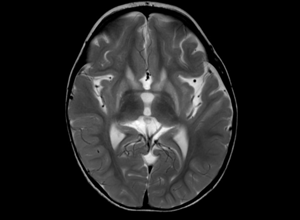
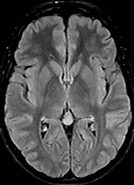
The most characteristic neuroradiological findings in Leigh syndrome are bilateral, symmetric focal hyperintensities in the basal ganglia, thalamus, substantia nigra, and brainstem nuclei at various levels on T2-weighted MRI. These high T2 signals on MRI reflect the spongiform changes and vacuolation in the affected brain structures . Often, the basal ganglia are affected before the brainstem. The upper brainstem followed by lower brainstem would be affected with the progression of the disease. Involvement of lower brainstem indicates advanced stage of the disease and the occurrence of respiratory failure and sudden death. In most patients the cerebral white matter is generally only involved in late stages of the disease. Occasionally, patients may have atypical neuroimaging features such as diffuse supratentorial leukodystrophy, unifocal or multifocal infarctions, diffuse or focal cortical atrophy, or predominant cerebellar atrophy

References:

10.2349/biij.6.1.e6

Crossed cerebellar diaschisis

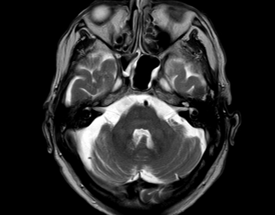
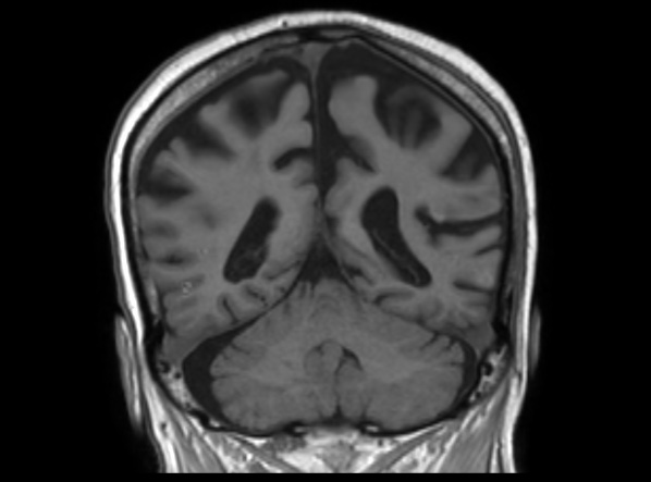
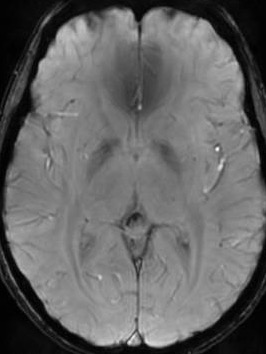
Crossed cerebellar diaschisis refers to a depression in function, metabolism, and perfusion affecting a cerebellar hemisphere occurring as a result of a contralateral focal supratentorial lesion, classically an infarct. 

Diaschisis in the brain, by definition, refers to an acute inhibition of function and metabolism produced by a focal disturbance in a portion of the brain at a distance from the original site of injury but connected via white matter tracts . Although initially defined as caused by an acute lesion, many sources in the literature employ a more liberal definition and refer to it being related to a lesion of any temporal duration .

When affecting the cerebellum, it is a well-recognized phenomenon following cerebral infarction , although it can be a sequela of any significant supratentorial lesion. The most likely mechanism is thought to be interruption of cortico-ponto-cerebellar white matter tracts, which then results in deafferentation and hypometabolism of the contralateral cerebellar hemisphere.

Reference:
doi:10.4103/0976-3147.102588

Infected thyroglossal duct cyst

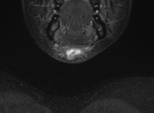
Presence of internal high attenuation, internal debris, and septations generally correlates with prior infection. In active or recent infection, patients may complain of tenderness at the site of a rapidly growing neck mass. Subsequent imaging reveals a thick-walled cyst with rim enhancement and inflammatory changes of the surrounding subcutaneous tissues.

Reference:
Patel, S., Bhatt, A.A. Thyroglossal duct pathology and mimics. Insights Imaging 10, 12 (2019). https://doi.org/10.1186/s13244-019-0694-x

Dacryocystocele

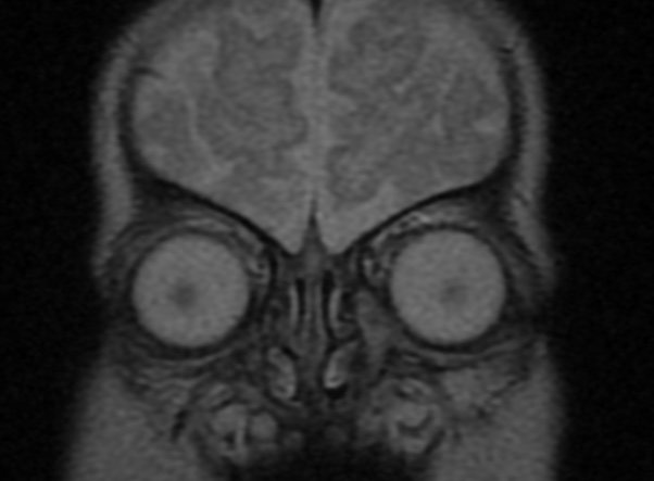
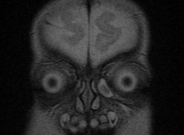
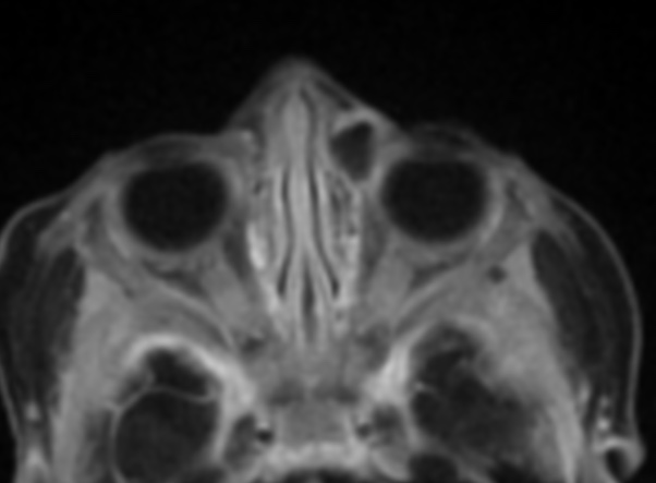
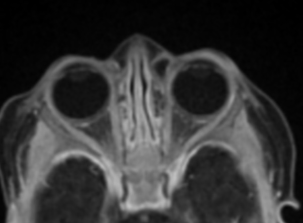
Congenital dacryocystocele is an uncommon consequence of congenital nasolacrimal duct obstruction: it is believed to occur as a result of a concomitant upper obstruction of the Rosenmuller valve and lower obstruction of the Hasner valve.This causes accumulation of fluid in the drainage system: the sac is initially filled with mucoid material with a grey-blue cystic swelling just below the medial canthus. Secondary dacryocystitis frequently develops within days or weeks

Fourth ventricle neurocysticercosis

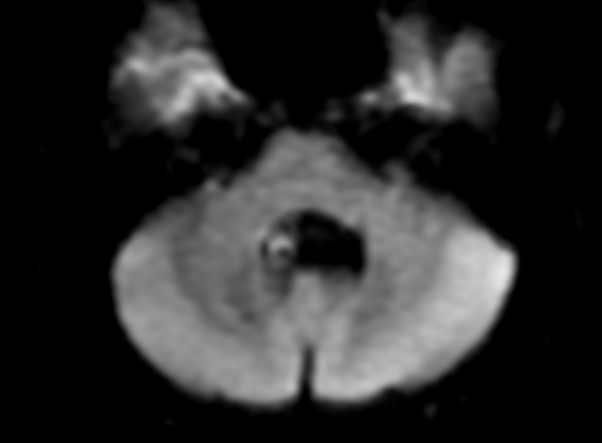
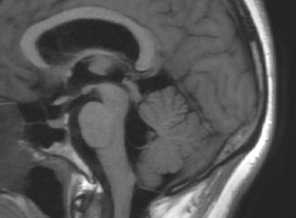
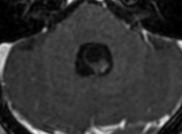
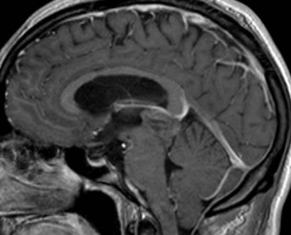
Extraparenchymal disease is known to be associated with more severe complications, such as hydrocephalus, arachnoiditis, and ventriculitis,resulting in a worse overall outcome. Intraventricular cysts are most found in the fourth ventricle (43%–70%) followed by the lateral (11%–43%) and third (1%–29%) ventricles with a minority in the aqueduct (7%–9%).Hydrocephalus is frequently present in intraventricular NCC cases due to mechanical obstruction of CSF flow or associated arachnoiditis.

Reference:

doi: 10.4103/sni.sni_218_18

lingual thyroid ectopia

Ectopic localization of the thyroid gland is an embryologic anomaly that occurs in the process of thyroid migration. When imaging is needed, MRI is one of the imaging modalities that can be used to visualize lingual thyroid. Some potential MRI findings associated with lingual thyroid may include:

  1. Hyperintense signal on T2-weighted images: Lingual thyroid tissue may appear hyperintense (bright) on T2-weighted MRI images, indicating increased water content.
  2. Isointense or hypointense signal on T1-weighted images: Lingual thyroid tissue may appear isointense (similar intensity) or hypointense (darker) on T1-weighted MRI images, compared to surrounding muscle tissue.
  3. Abnormal location: MRI may show the presence of thyroid tissue in an abnormal location within the tongue, typically located at the base or posterior aspect of the tongue.
  4. Absence or atrophy of thyroid tissue in the normal location: MRI may show an absence or atrophy of the thyroid gland in its normal location in the neck, which may suggest compensatory hypertrophy of the lingual thyroid.

Pineal parenchyma tumour of intermediate differentiation

Pineal parenchyma tumour of intermediate differentiation (PPTID) accounts for up to 20 % of all pineal parenchymal tumours. They are characterised by diffuse sheets or lobules of cells similar to pineocytomas and pineoblastomas with mild to moderate nuclear atypia and low to moderate mitotic activity. PPTID are of intermediate-grade malignancy and are classified as WHO grade II or III neoplasm. The peak prevalence is during adulthood; however, they can occur at all ages and have a female predilection. The 5-year survival is 39–79 %, which is between the outcomes of pineocytoma and pineoblastoma .

No specific MRI findings distinguish PPTID from pineocytomas or pineoblastomas. Generally, PPTID are lobulated, vascular pineal region masses that can extend into adjacent structures such as the ventricles or thalami. Owing to high cellularity, PPTID are usually hyperdense on CT scans and can demonstrate peripheral exploded calcifications. On MRI, these tumours are heterogeneously hypointense on T1 weighted and heterogeneously hyperintense on T2 weighted images. Cystic areas can be seen within the tumour as well. Heterogeneous enhancement is typical. Hydrocephalus is often seen owing to mass effect on the tectum. Rarer complications include intracranial dissemination and cerebrospinal fluid spread to the spine.

References:

10.1259/bjrcr.20150371

10.1007/s13244-013-0248-6

Pineal cysts

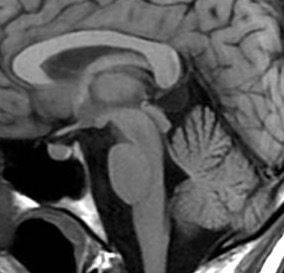
Pineal cysts are frequently asymptomatic, small sized, unilocular, benign pineal lesions which do not show size change. They are generally made of three layers: fibrocollagen layer at the outside, pineal parenchymal layer which may have calcium deposits at the middle and hypocellular glial tissue layer which may have hemosiderin inside. PCs may develop as secondary to focal degeneration of pineal gland or distension of pineal diverticulum remnant. Unilocular, smooth edged, round or ovoid shaped cysts which have homogenous interior signal feature and rim-shaped contrast-enhancement with less than 2 mm wall thickness on MRI are referred as typical PC.

Their frequency is higher in women and adults, and their sizes are not associated with gender or age. Great majority of them are isointense with CSF on T1 and T2A series. On FLAIR sequence, they are hyperintense compared to CSF, and they may be smoothly contoured, unilocular or multilocular. Typical ones may have contrast-enhancement in peripheral rim style, while multilocular ones may have septal contrast-enhancement.

Reference:

10.4329/wjr.v10.i7.65