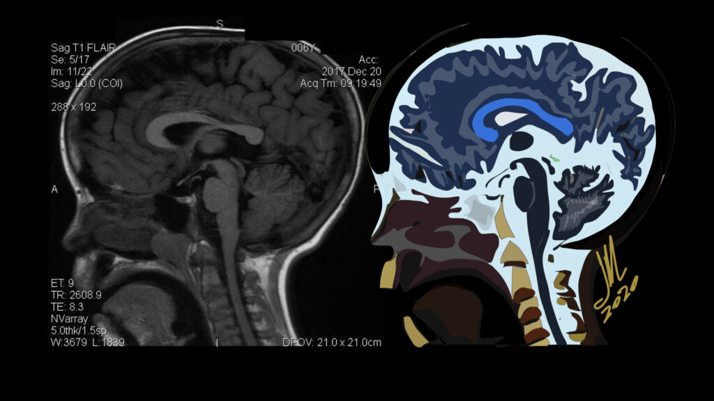
Acromegaly

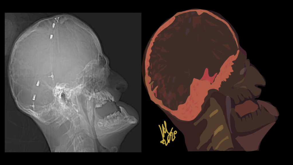
Characteristic maxillofacial findings of acromegaly include thickening of the calvaria, more pronounced in the inner table, enlarged paranasal sinuses, most often the frontal sinus, resulting in frontal bossing, and enlargement of the mandible, which may lead to prognathism and gaps between the teeth. Diffuse and symmetric enlargement of the extraocular muscles has been described as part of the generalized organomegaly associated with acromegaly.

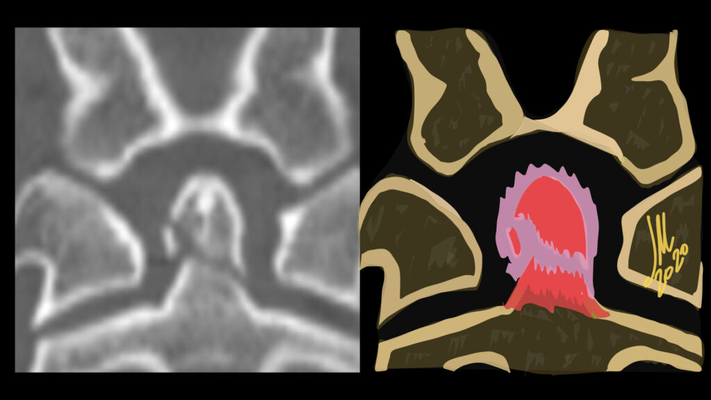
Successful surgical treatment of functional pituitary microadenoma largely relies on accurate identification of the tumor within the sella turcica.These relatively small tumors represent a challenge to both neuroradiologists and neurosurgeons in locating them, resulting in a greater potential for insufficient treatment of the lesion.
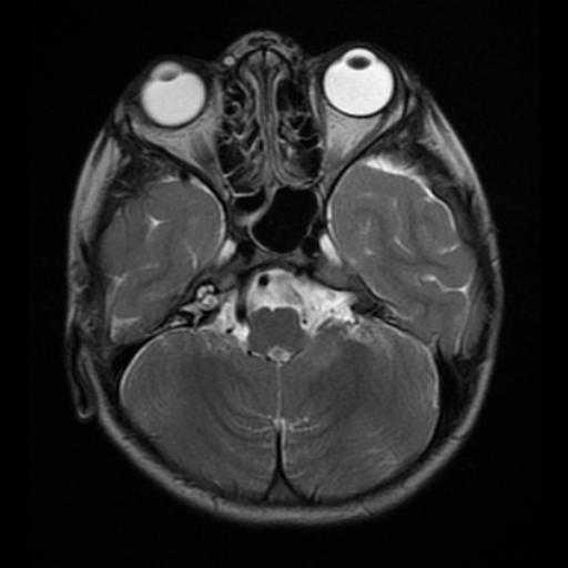
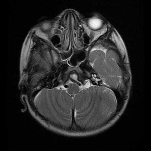
The criterion standard technique used for lesion localization is MR imaging. Dynamic contrast-enhanced technique is often applied on MR imaging to overcome this issue, scanning time usually required to obtain each dynamic phase ranges from 20 to 30 seconds or is shortened into 12–20 seconds in some cases, but it is not possible to obtain a gapless 3D image. The most suitable time phase to obtain sufficient contrast between an adenoma and the normal pituitary gland is 45–60 seconds after contrast agent injection. This adenoma-normal pituitary gland contrast will rapidly diminish within the subsequent 30 seconds. Both spatial and temporal resolution must, therefore, be sufficiently high to visualize the presence of the adenoma.

Acromegaly results from excessive production of growth hormone, most commonly in patients with pituitary adenoma >95% of cases, resulting in the overproduction of insulinlike growth factor 1. Acromegaly is a multisystem disease characterized by somatic overgrowth of osseous structures such as the hands, feet, skull, mandible, and nose. Soft-tissue thickening of the skin and vocal cords and enlargement of internal organs including the heart and kidneys also can be seen. Overproduction of growth hormone before epiphyseal closure leads to linear overgrowth and gigantism
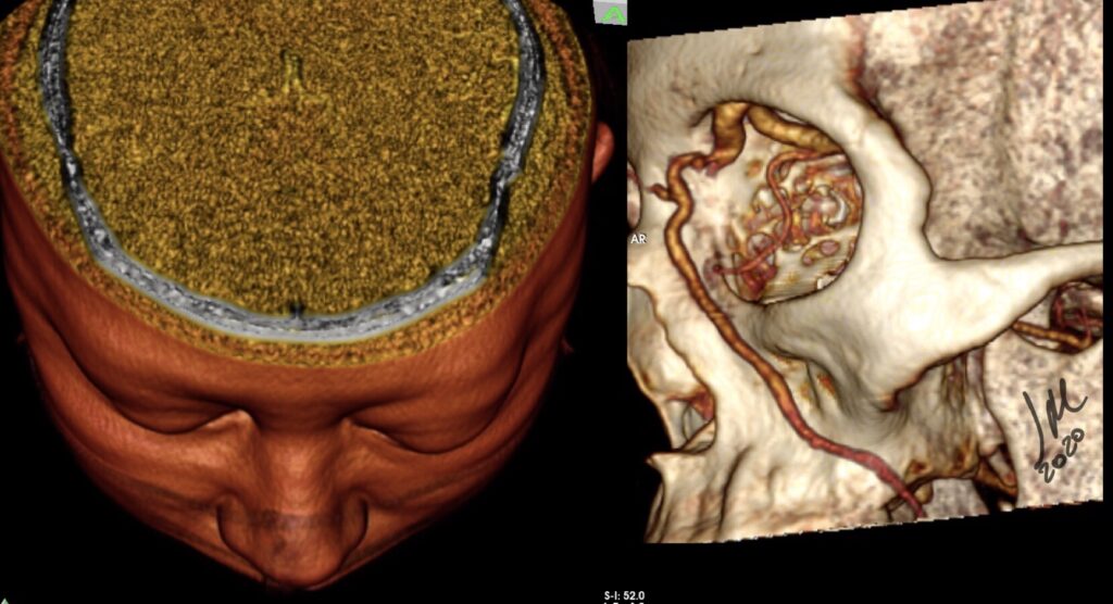
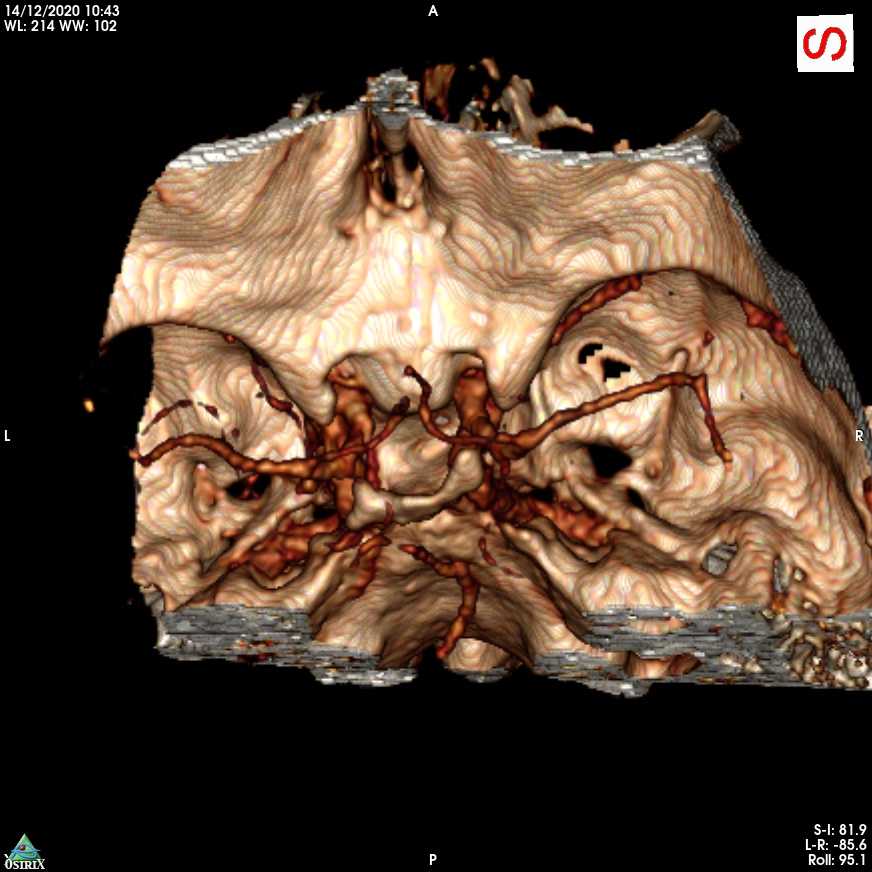
Spontaneous Carotid cavernous fistula

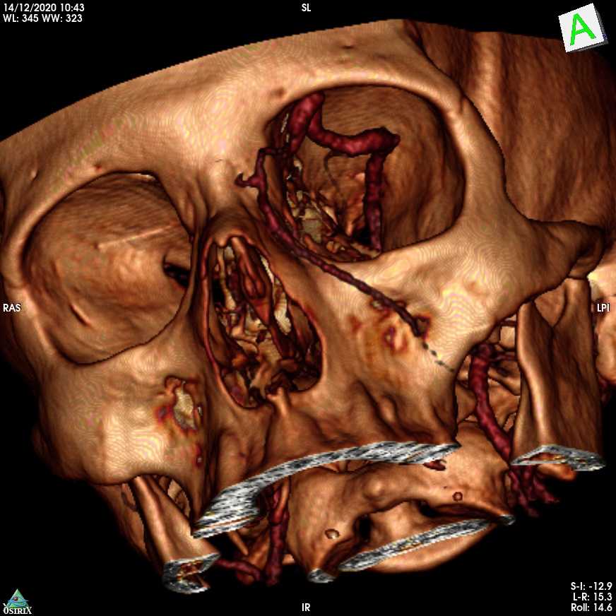
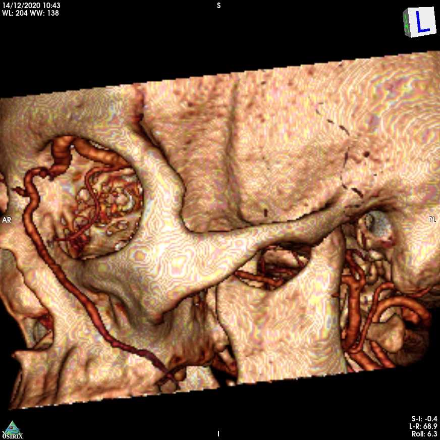
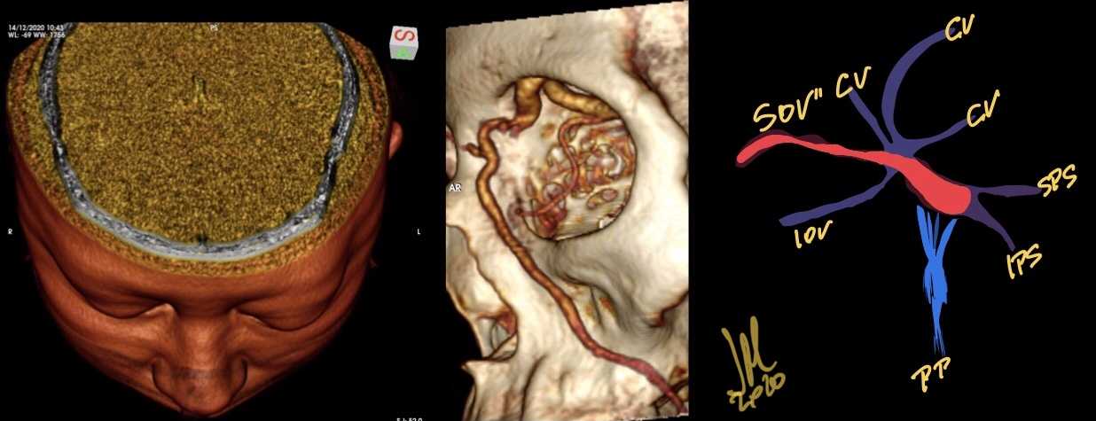
Pulsating exophthalmos, bruit, and conjunctival chemosis have long been regarded as the three classical symptoms of carotid cavernous fistula. If the main venous drainage is via the superior ophthalmic vein, this triad will probably be present; if not, the triad is likely to be absent or incomplete.

The symptoms of patients with spontaneous CCF are commonly milder than those in traumatic CCF cases. This difference is attributable to the difference in the volume of blood flow into the cavernous sinus. In addition, the specific venous drainage from the cavernous sinus is important.

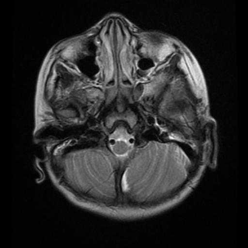
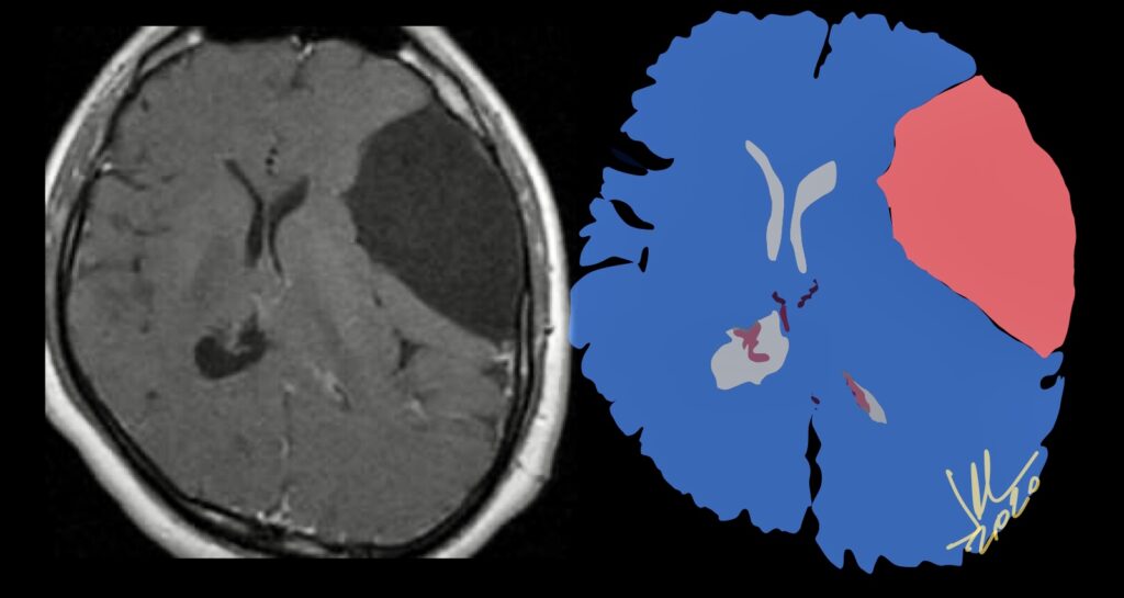
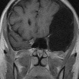
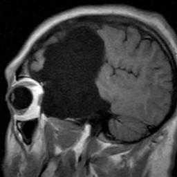
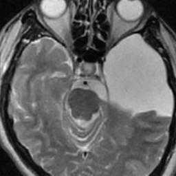
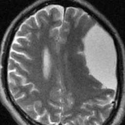
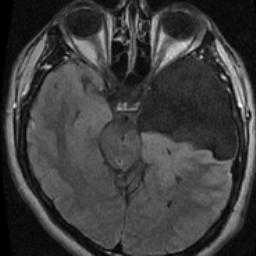
Galassi type III-Arachnoid Cyst.

GALASSI CLASSIFICATION OF MIDDLE TEMPORAL FOSSA IACS

MTF ACs account for more than half of all ACs . The widely used Galassi Classification (1982) provides a schematic radiological classification of these lesions. There are three types of MTF IACs based on size and degree of mass effect.

Type I cysts are characterized by the following: lens-shaped, anterior tip of the MTF, freely communicate with CSF/surrounding subarachnoid space on MR-cine/CT-ventriculography, they rarely require surgery.

Type II cysts are characterized by intermediate size, more rectangular morphology, extend into the Sylvian fissure, have variable communication with CSF pathways, and exert local mass effect on the temporal lobe; they occasionally and sometimes require surgery.

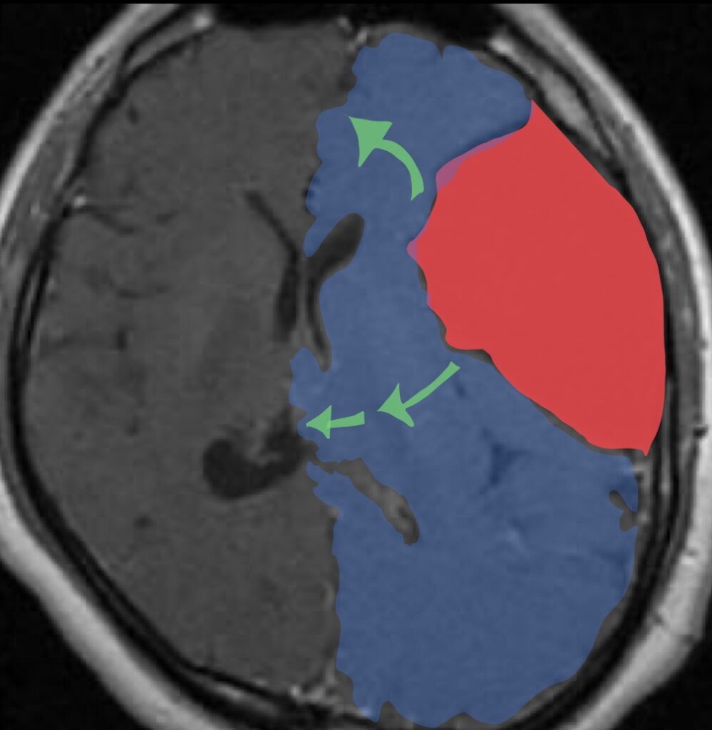
Type III cysts constitute the largest group, extend the full length of the Sylvian fissure, exert significant mass effect (often with midline shift), and do not communicate with the subarachnoid space; these usually require surgery.

There is no Class I evidence regarding the optimal treatment of ACs. We must, therefore, weigh the risks versus benefits of conservative versus surgical treatment of these lesions in symptomatic patients on an individual patient basis. 

Choanal Atresia

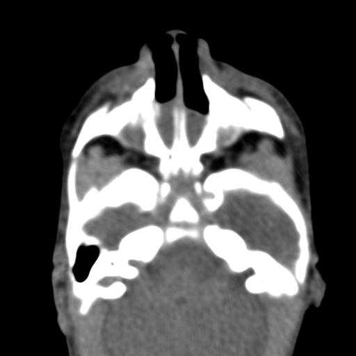
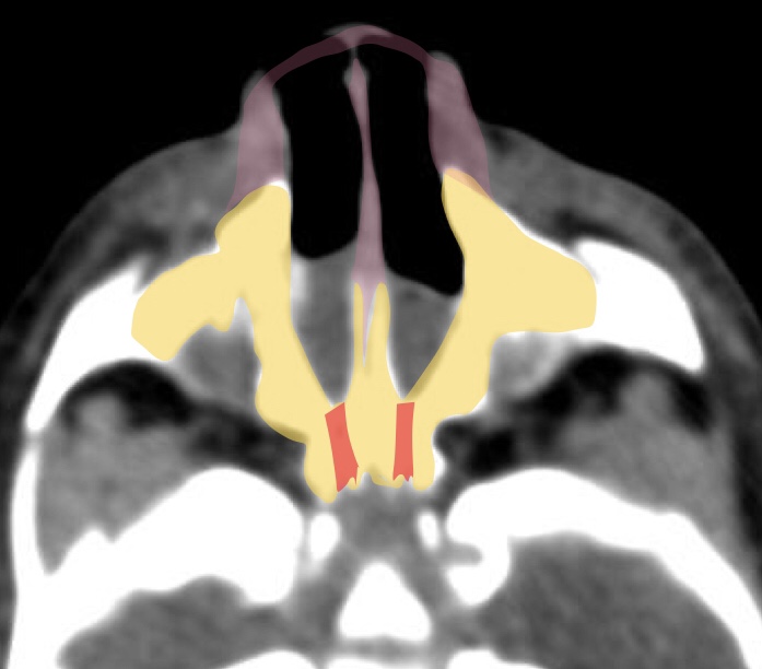
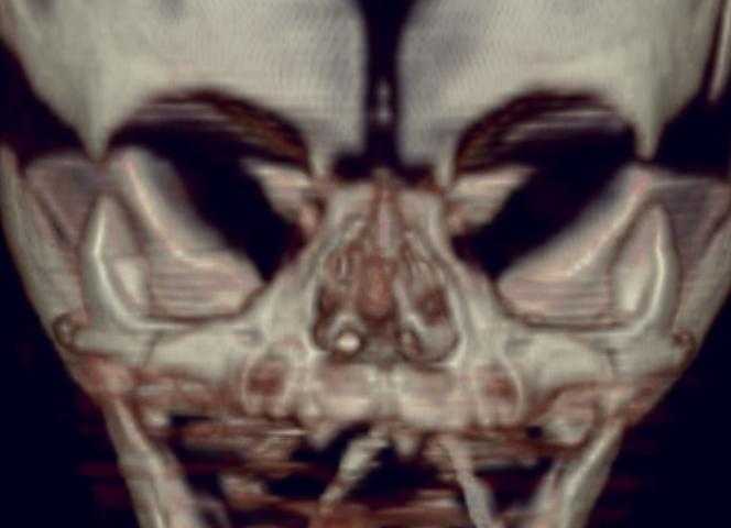
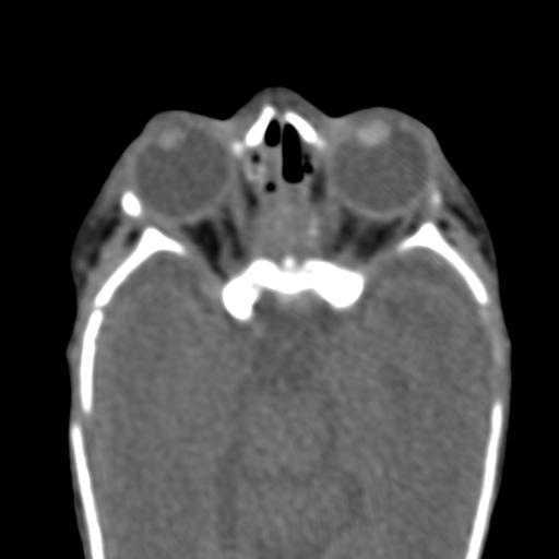
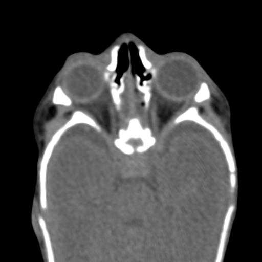
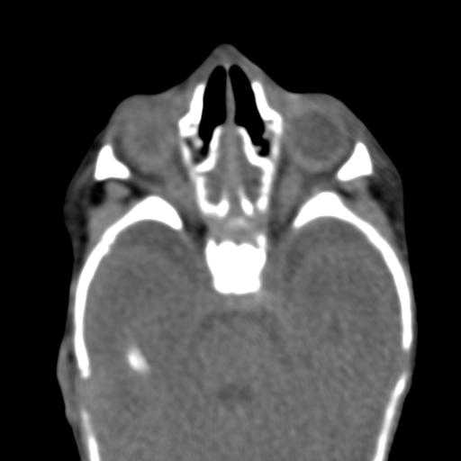
Choanal atresia is an uncommon developmental defect characterized by lack of communication between the nasal cavity and the nasopharynx. This plate may be bony (70-90%), membranous (10%), or both. The atresia may involve one or both nasal cavities, and other congenital anomalies may be associated with it. If bilateral choanal atresia is present at birth, it requires immediate diagnosis and intervention to permit respiration. Unilateral choanal atresia is usually diagnosed at a later stage.

CT in the axial plane was extremely valuable in demonstrating choanal atresia. The configuration of the nasal cavity is displayed in its entire length and it is possible to visualize stenosis and septal deviation and to differentiate between the bony and membranous components.

Other congenital anomalies such as craniofacial cleft palate, Treacher Collins syndrome, and cardiovascular and abdominal malformations have been reported in 20%-50% of cases of choanal atresia

Lymphangioma

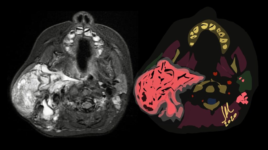
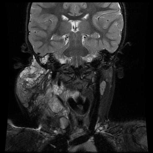
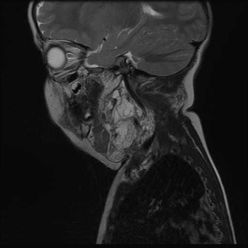
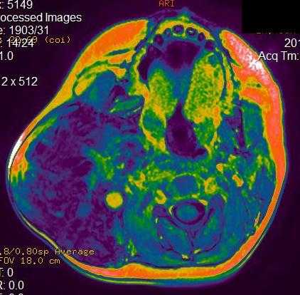
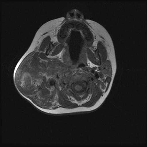
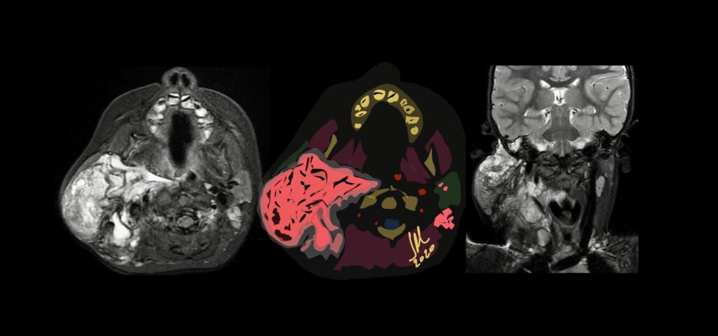
Lymphangiomas are rare congenital benign lesions occurring mainly in the head, neck and oral cavity frequently present at birth. Cystic hygromas, however, account for approximately 90% of the lymphangiomas in the head and neck region. Other common sites, outside the head and neck, include the axilla, shoulder, chest wall, mediastinum, abdominal wall, and thigh. They have no predilection for sex or race, and they have no malignant potential. Typical cystic hygromas cause no symptoms unless they enlarge in size or surround or invade adjacent normal anatomic structures. Cystic hygromas may cause symptoms such as feeding problems or breathing difficulties. Cystic hygromas are multilobulated, thin-wall, lymph-containing sacs. The fluid within the sacs is usually clear or amber colored, although occasionally it could be turbid or hemorrhagic.

MR imaging is useful to confirm the diagnosis and define the extension of the cystic lesions and their relationship to adjacent structures. A diagnostic dilemma may occur if a cystic hygroma containing a suspicious vascular anomaly is seen in the lower neck or upper mediastinum. In this case, the vascular malformation should be differentiated from a thoracic duct aneurysm.  

They consist in localized centres of abnormal development of the lymphatic system. Three theories have been proposed to explain the origin of this abnormality. The first suggests that a blockage or arrest of normal growth of the primitive lymph channels occurs during embryogenesis, the second that the primitive lymphatic sac does not reach the venous system, while the third advances the hypothesis that, during embryogenesis, lymphatic tissue lays in the wrong area

Neurovascular Compression of Facial Nerve

Neurovascular compression syndrome (NVCS) is defined as a direct contact with mechanical irritation of cranial nerves by blood vessels.  By far the cases of neurovascular contact are clinically asymptomatic

NVCS can occur either at the root entry-exit zone 10%–90%,  adjacent cisternal segment 60%, Transitional zone 22%.

Microvascular decompression is highly effective in HFS and symptoms disappear after an operation in 90%–95% of cases; however, recurrence is seen in up to 25% of patients.

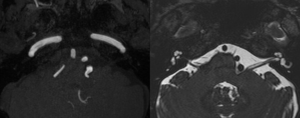
high-resolution 3D T2-weighted imaging with 3D time-of-flight angiography and 3D T1-weighted gadolinium-enhanced sequences is considered the standard of reference for the detection of neurovascular compression 

The transition zone between central and peripheral myelin is an anatomic area with increased mechanical vulnerability, which is of particular interest in the context of symptomatic NVCS. The transitional zone look to be the more relevant and vulnerable anatomic structure, and it is not always located in the same position as the root entrance-exit zone (TZ). Exact anatomic knowledge of the position and morphology of the TZ is of fundamental importance for the interpretation of neuroimaging findings in suspected NVCS. TZ of VII CN to be about <1mm to REZ.

To remember TZ location from REZ:

CN V = 2mm. CN VIII = 1mm. CN VIII= 10mm. CN IX= 15mm

STIR Sequence for Acute Type II-III

Type III

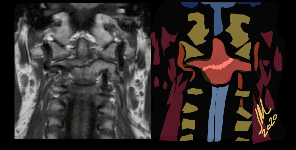
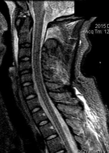
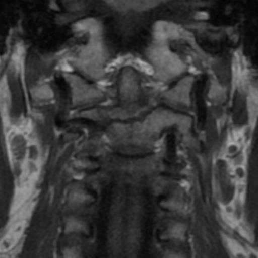
Because MR imaging and the STIR sequence are used to determine fracture acuity and, therefore, treatment recommendations, it is important that we understand the limitations of this sequence.The STIR sequence is relied on to reveal marrow edema related to an acute fracture. In addition to the increased detection of soft-tissue injury, the STIR sequence is exquisitely sensitive to bone marrow pathology. This feature may be useful in identifying the acuity of a fracture. The STIR sequence, while sensitive for the detection of acute type II-III odontoid fracture in patients younger than 50 years, is significantly less sensitive in older patients, particularly those with osteopenia. In older patients The combination of the vascular watershed and the lack of a periosteal blood supply contributes to overall poor vascularity in the odontoid, which contributes to the absence of bone marrow edema and, subsequently, the absence of STIR hyperintensity in the setting of acute fracture in the older population. Evaluating all the sequences is imperative and the combination of the t1 sequence in the coronal and sagittal plane with the stir sagittal and coronal sequence is very useful.

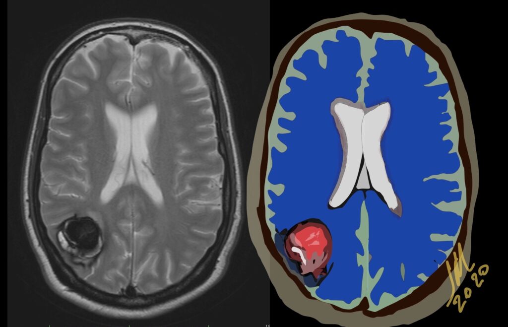
Atypical hemorrhage due to Zabramski type V Cavernoma

Type V indicate parts of the actual CCMs that are visible in the center of the hemorrhage; however, the CCM is not fully distinguishable from hemorrhage. Type 5 has a high hemorrhage risk of 24%

Hemorrhagic events particularly those found in relatively young patients ought to be characterized further, and cavernous angioma should be considered among the possible etiologies. Also because of their propensity to act as an epileptogenic focus CMs should always be considered in the workup of a patient with a seizure disorder, especially if the patient is aged 20-40 years.

If a recent bleed has occurred then surrounding edema or mass effect may be present. The lesions show a variable pattern of enhancement. Contrast-enhanced images help delineate any potential associated developmental venous anomalies. This is critical for preoperative surgical planning as the un-deliberate resection of DVAs may compromise normal cortical venous drainage and thus lead to brain venous infarction. Angiographically these lesions are occult and usually show nonspecific findings like capillary blush or early draining vein

Odontoid type 2 fracture

Fracture through the base of the dens. Type II odontoid fractures are potentially unstable with subsequent myelopathy and spinal injury risk. Higher risk of nonunion with comminuted fracture, age > 50 or more than 5 mm displacement.

Odontoid fracture is a very common cervical injury, accounting approximately for 9%–15% of all cervical fractures. These injuries result from an hyperextension or hyperflexion mechanism of the cervical spine in low-energy impacts such as falls for elderly patients or in high-energy impacts such as motor vehicles accidents for young people.
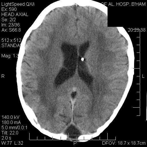
The classification scheme of odontoid fractures described by Anderson and D’Alonzo is the one most commonly used. However there is a new classification by Grauer who proposed a better difference between type 2 -3.

Cervical spine trauma is most commonly evaluated with CT with MPR reconstructions 1-3mm.

Hutchinson-Gilford Progeria Syndrome

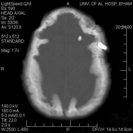
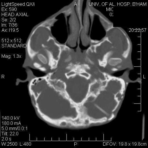
Hutchinson-Gilford Progeria Syndrome (HGPS) is a rare syndrome of segmental premature aging. Children appear healthy at birth and within the first year of life develop severe failure to thrive with sclerodermatous skin changes. Classic craniofacial features develop with age and underlying disease culminates in death from myocardial infarction or stroke.
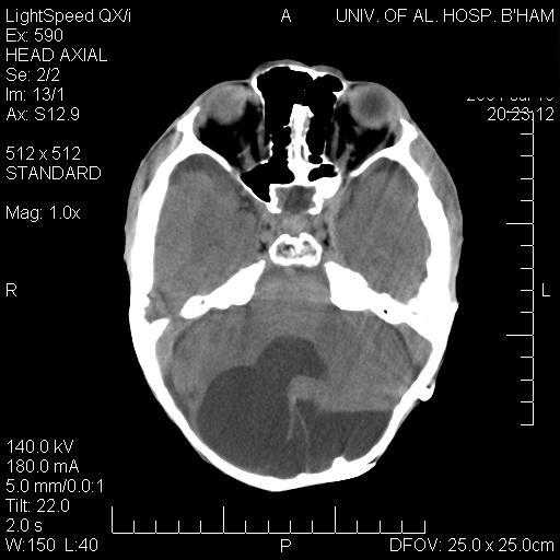
Thinning and mottling of the calvarium, prominent vascular markings, delayed closure of the anterior and posterior fontanels, widening of calvarial sutures, and a J-shaped sella, optic nerve kinking and hypotelorism, short mandibular rami in combination with flattened mandibular condyles are observed.