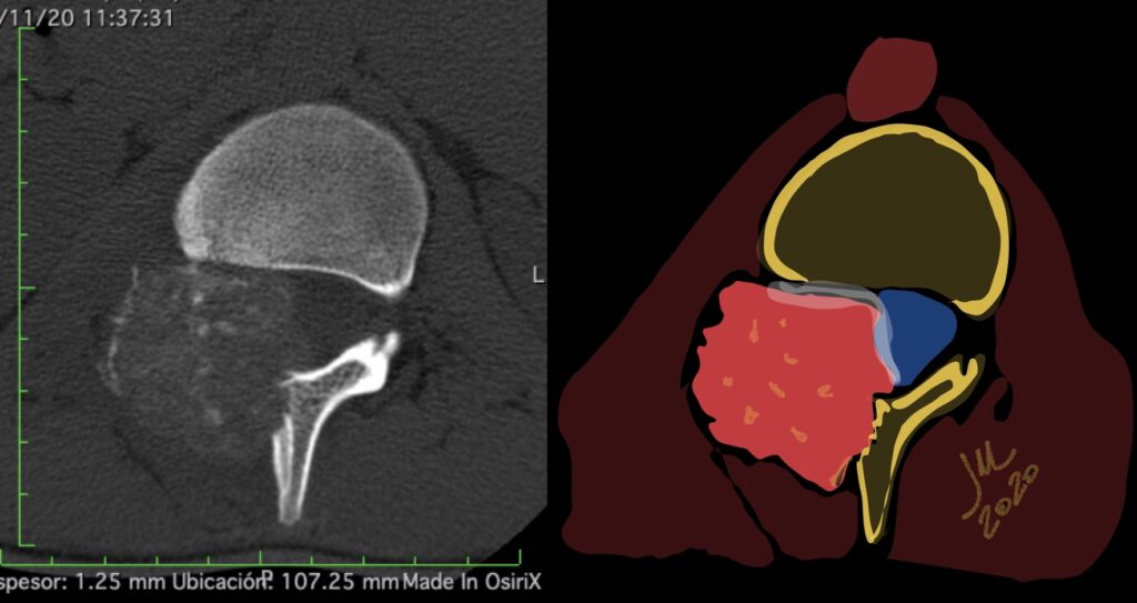
OBs are very rare and constitute about 1% of all primary bony tumors. These tumors tend to involve long bones and vertebral column. OBs are rare primary bony tumors constituting only 1% of all cases.
In 1984, a borderline osteoblastic tumor entity called “aggressive osteoblastoma” was introduced by Dorfman and Weiss and was found to be associated with a high recurrence rate and potential for malignant transformation. Despite being benign tumors, osteoblastomas often cause pronounced bone destruction, soft tissue infiltration, and epidural extension. They usually behave aggressively with extensive uncontrollable local recurrence, and even malignant transformation with metastatic disease has been reported
CT appearance of spinal osteoblastoma is a lytic expansile lesion with a shell of sclerosis, and soft tissue masses can be observed around the lesion.
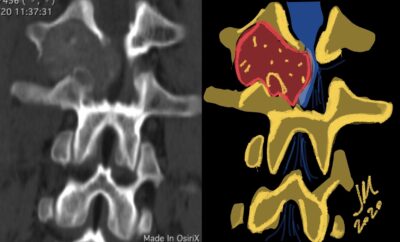
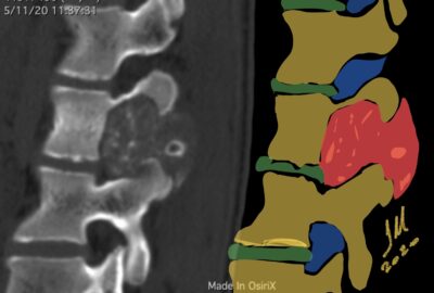
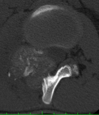
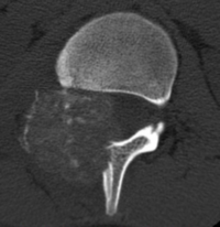
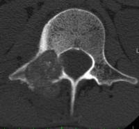
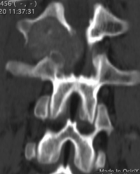
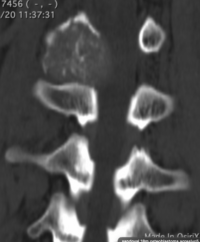
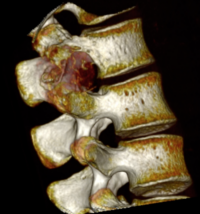
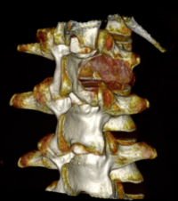
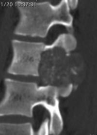
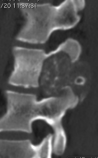
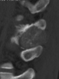
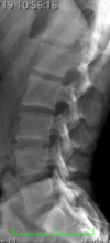
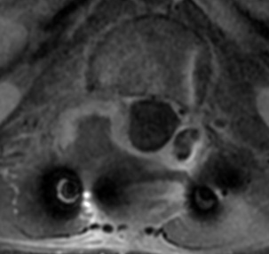
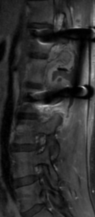
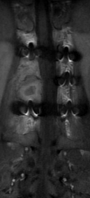
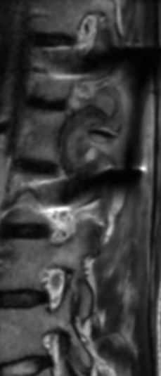
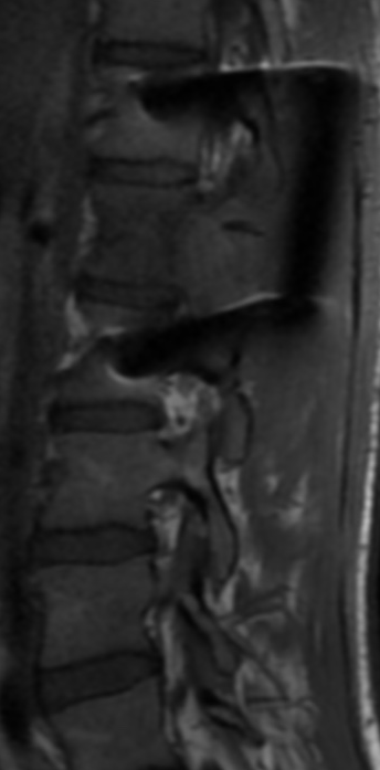
The imaging features of spinal osteoblastoma on MRI are considered nonspecific. These lesions often display a low to isointense signal on T1-weighted MRI, whereas T2-weighted MRI demonstrates an intermediate- to high-intensity signal due to matrix calcification and intense enhancement representing the highly vascular nature of such lesions
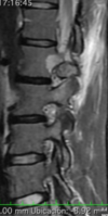
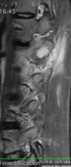
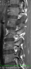
Notably, however, this radiographic appearance may lead to overestimation of the limits of the tumor and can be somewhat confusing to orthopedic surgeons and radiologists, who may interpret these tumors as other clinical entities such as Ewing’s sarcoma or lymphoma
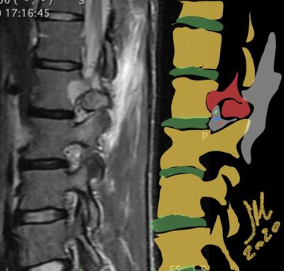
flare phenomenon” was described by Crim et al. in 1990. Specifically, these neoplasms often cause a diffuse, reactive prostaglandin (PG) – mediated inflammatory infiltrate within adjacent vertebrae surrounding paraspinal soft tissues. In contrast, for aggressive osteoblastoma in the spine, the tumor may be an expansile lesion with a multitude of small calcifications, a prominently clerotic rim, and paravertebral and epidural extensions and may also radiographically mimic aneurysmal bone cysts, osteosarcomas, or bone metastases.
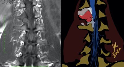
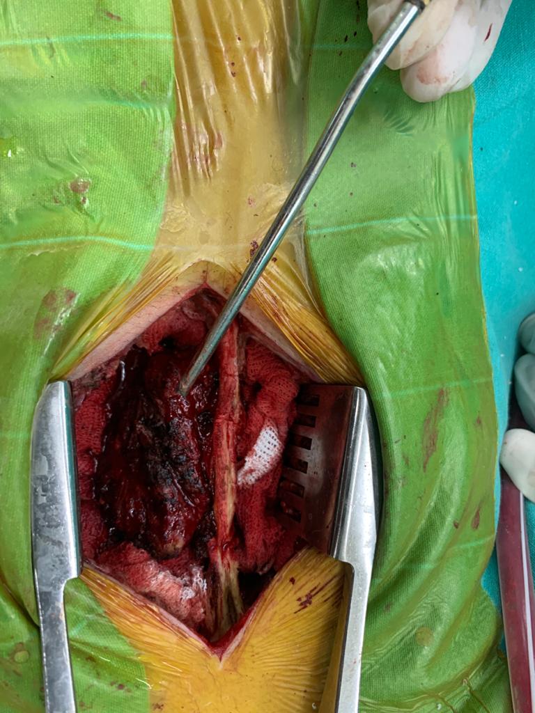

Another essential diagnostic tool is bone scintigraphy, which is the most sensitive radiographic scan described in the literature for osteoblastoma and typically shows increased radiotracer uptake, indicating increased osseous turnover